Review
. 2023 Oct 12;20(1):42.
doi: 10.1186/s12014-023-09431-y.

Redefining serological diagnostics with immunoaffinity proteomics

Affiliations
Review

Redefining serological diagnostics with immunoaffinity proteomics

Jonathan Walter et al. Clin Proteomics. .

Abstract

Serological diagnostics is generally defined as the detection of specific human immunoglobulins developed against viral, bacterial, or parasitic diseases. Serological tests facilitate the detection of past infections, evaluate immune status, and provide prognostic information. Serological assays were traditionally implemented as indirect immunoassays, and their design has not changed for decades. The advantages of straightforward setup and manufacturing, analytical sensitivity and specificity, affordability, and high-throughput measurements were accompanied by limitations such as semi-quantitative measurements, lack of universal reference standards, potential cross-reactivity, and challenges with multiplexing the complete panel of human immunoglobulin isotypes and subclasses. Redesign of conventional serological tests to include multiplex quantification of immunoglobulin isotypes and subclasses, utilize universal reference standards, and minimize cross-reactivity and non-specific binding will facilitate the development of assays with higher diagnostic specificity. Improved serological assays with higher diagnostic specificity will enable screenings of asymptomatic populations and may provide earlier detection of infectious diseases, autoimmune disorders, and cancer. In this review, we present the major clinical needs for serological diagnostics, overview conventional immunoassay detection techniques, present the emerging immunoassay detection technologies, and discuss in detail the advantages and limitations of mass spectrometry and immunoaffinity proteomics for serological diagnostics. Finally, we explore the design of novel immunoaffinity-proteomic assays to evaluate cell-mediated immunity and advance the sequencing of clinically relevant immunoglobulins.

Keywords: Immunoassays.; Immunoglobulins; Mass spectrometry; Proteomics; Serological diagnostics.


Conflict of interest statement

The authors declare no competing interests.

Figures

Fig. 1
Fig. 1
Serological immunoassays with conventional detection techniques. (A) Colorimetric indirect ELISA: antigens of interest are immobilized on the microplate surface and incubated with diluted blood serum samples. Specific human antibodies (blue) are captured, and non-specific antibodies and proteins are removed with microplate washing. Enzyme-conjugated secondary anti-human antibodies (green) oxidize the substrate (yellow), and the absorbance of the product is measured by a spectrophotometer. Relative antibody titers are determined by the highest dilution of a positive blood serum sample that provided a positive result. (B) Lateral flow immunoassay: Specific antibodies in patient samples bind to an antigen immobilized on colloidal gold nanoparticles on a sample pad (S). Capillary flow transfers complexes to the conjugation pad, where the complexes interact with the immobilized anti-human secondary antibodies, aggregate, and precipitate at the test line (T). Precipitation of gold nanoparticles results in color change (red stripe). As a control for test completion, rabbit antibodies conjugated to gold nanoparticles travel along through the T region, interact with the goat-anti-rabbit antibodies at the control line C, precipitate, and result in color change. (C) Multiplex particle-based flow cytometry: Each antigen is conjugated to a bead of a unique “color” which is predetermined by a unique combination of ten infrared dyes at different concentrations. Beads are mixed and incubated with serum, and the antigens capture corresponding human antibodies. Secondary anti-human IgG antibodies are conjugated to a fluorescent dye (green) used for quantification. Particle-based flow cytometry utilizes two different lasers to detect the bead identity (red laser) and signal intensity (green) of a single bead passing through the detection region. To map antibody isotypes and subclasses across multiple antigens, the analysis is repeated with the secondary antibodies specific for human IgM, IgA, or IgG1-4
Fig. 2
Fig. 2
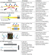
Emerging immunoassay detection technologies for serological diagnostics. (A) Surface plasmon resonance assays detect antibodies through their binding to antigens immobilized on gold surfaces. Specific binding results in plasmon resonance and changes in the refractive index. (B) A biolayer interferometry immunosorbent assay detects the shift in the wavelength of the reflected light upon antigen-specific antibody binding to the surface of a fiber optic sensor. (C) Electrochemical biosensors are transistors that detect antibody-antigen binding onto a gate electrode and measure changes in voltage across the source and drain electrodes. (D) Protein microarrays consist of numerous antigens that are printed onto the surface, enrich specific antibodies, and facilitate their relative quantification using fluorophore-labeled secondary antibodies. (E) Following affinity enrichment and trypsin digestion, MS facilitates highly specific quantification of peptides representing human immunoglobulin isotypes and subclasses
Fig. 3
Fig. 3
Serological diagnostics with immunoaffinity-targeted proteomics. (A) Specific antibodies are affinity enriched from the patient serum, denatured, and digested with trypsin, and the peptides are analyzed by targeted MS assays. (B) Following electrospray ionization (ESI), targeted MS assays, such as Selected Reaction Monitoring, enable the isolation of peptide ions of interest with the first quadrupole (Q1), their fragmentation in Q2, isolation of fragments of interest in Q3, and measurement of fragment ion intensities. (C) Peak areas of the “light” endogenous peptides and the corresponding spiked-in “heavy” isotope-labeled internal standards are used to calculate peptide ratios and absolute concentrations of the complete panel of the human immunoglobulin isotypes and subclasses
Fig. 4
Fig. 4
Challenges of characterization and sequencing of polyclonal antibodies by MS. Variable heavy (VH) and light (VL) chains of human immunoglobulins include semi-variable framework regions (FR1-4) and hypervariable CDRs, with CDR-H3 being the most diverse domain. Sequence logos present the experimental diversity of the matched heavy and light variable chains of 199 B-cell clones secreting anti-spike SARS-CoV-2 antibodies, based on reanalysis of data for a single convalescent donor [158]. The framework regions are encoded by the germline V- and J-genes, are minimally affected by affinity maturation, and include some well-conserved sequences (YYCAR, etc.) suitable as sequence tags for the hybrid de novo sequencing approaches
Fig. 5
Fig. 5
The proposed IA-MS workflow for in vitro measurements of pMHC-specific TCRs. (A) Immunopeptidome workflows facilitate the identification of MHC-specific peptides through the isolation of antigen-presenting cells, affinity enrichment of peptide-MHC (pMHC) complexes, dissociation of class I (9–10 aa) or class II (13–25 aa) MHC peptides, and de novo sequencing of MHC peptides by LC-MS. (B) Recombinant MHC I or MHC II proteins representing the patient-specific Human Leukocyte Antigen (HLA) variants are conjugated to magnetic particle-bound streptavidin tetramers and incubated with the synthetic MHC I or MHC II peptides previously identified with immunopeptidome workflows. Numerous pMHC complexes need to be prepared. (C) CD8 + cytotoxic T lymphocytes (MHC I) and CD4 + helper T lymphocytes (MHC II) are isolated from the patient’s blood and lysed to release soluble TCRs (α-β1 or α-β2). (D) T lymphocyte lysates are incubated with the individual pMHC complexes, and endogenous pMHC-specific TCRs are enriched and covalently cross-linked to pMHCs to retain low-affinity interactions. Following trypsin digestion, unique peptides of α constant (TRAC_HUMAN), β1 constant (TRBC1_HUMAN), and β2 constant (TRBC2_HUMAN) chains are quantified by LC-SRM. Corresponding heavy isotope labeled peptide internal standards (IS) facilitate accurate relative or absolute quantification

Similar articles

Cited by

References

    1. Wassermann, Av Bruck CGv. Experimentelle Studien über die Wirkung von Tuberkelbacillen-Präparaten auf den tuberculös erkrankten Organismus. Dtsch Med Wochenschr. 1906;32:449–54.
    1. Allard-Chamard H, Boire G. Serologic diagnosis of rheumatoid arthritis. Clin Lab Med. 2019;39(4):525–37. - PubMed
    1. Lindfors K, Ciacci C, Kurppa K, Lundin KEA, Makharia GK, Mearin ML, et al. Coeliac disease. Nat Reviews Disease Primers. 2019;5(1):3. - PubMed
    1. Yang W-L, Lu Z, Guo J, Fellman BM, Ning J, Lu KH, et al. Human epididymis protein 4 antigen-autoantibody complexes complement cancer antigen 125 for detecting early-stage ovarian cancer. Cancer. 2020;126(4):725–36. - PMC - PubMed
    1. Brownstein NC, Chen YA. Predictive values, uncertainty, and interpretation of serology tests for the novel coronavirus. Sci Rep. 2021;11(1):5491. - PMC - PubMed

LinkOut - more resources