. 2023 Oct 11;23(1):746.
doi: 10.1186/s12903-023-03399-9.

Positional transfer accuracy of titanium base implant abutment provided by two different scan body designs: an invitro study

Affiliations

Positional transfer accuracy of titanium base implant abutment provided by two different scan body designs: an invitro study

Rania E Ramadan et al. BMC Oral Health. .

Abstract

Background: The variabilities in design and material of scan bodies have a major role in the positional transfer accuracy of implants. The purpose of this invitro study was to compare the 3D transfer accuracy (trueness and precision) of titanium base (TB) abutment position provided by 2 different scan bodies: one-piece scan body (SB) in comparison to two-piece healing abutment and scan peg (HA-SP).

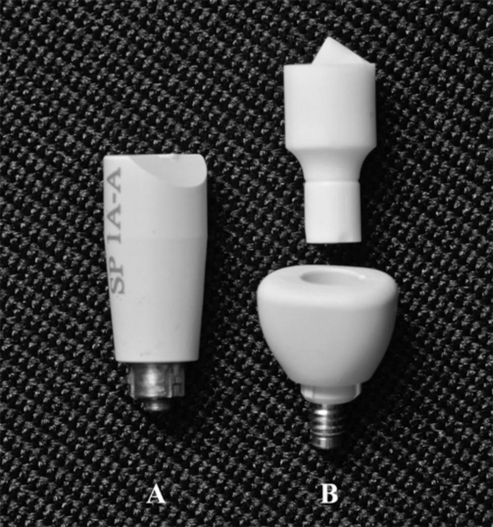
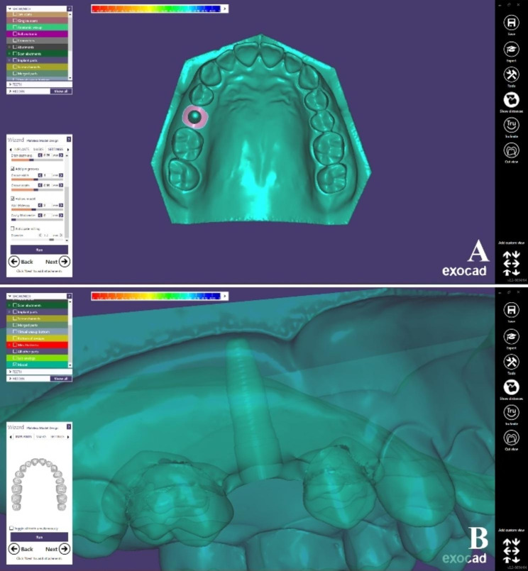
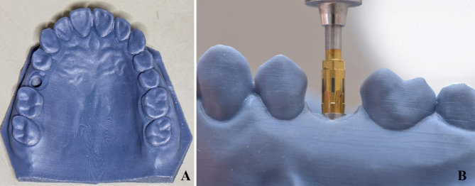
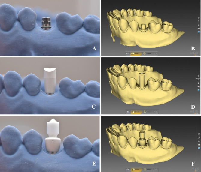
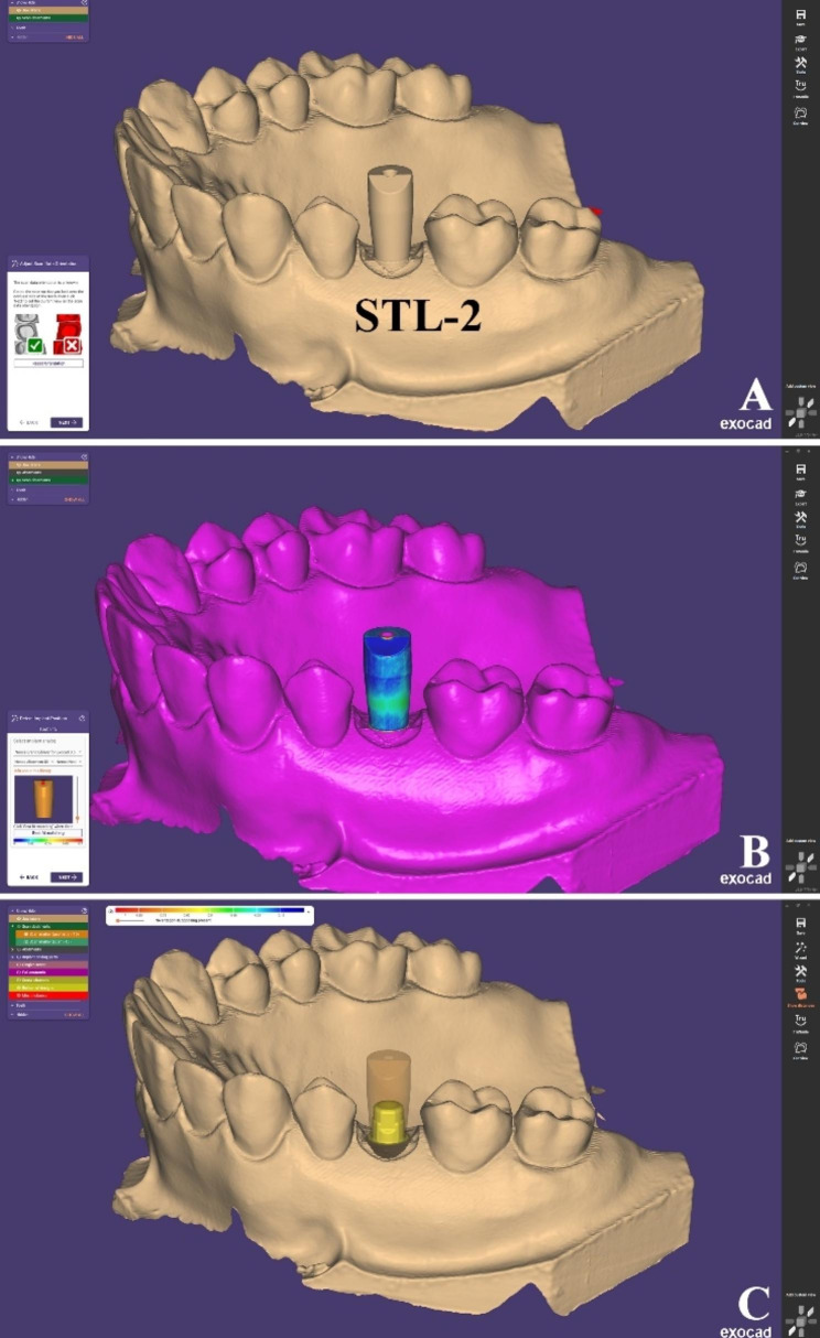
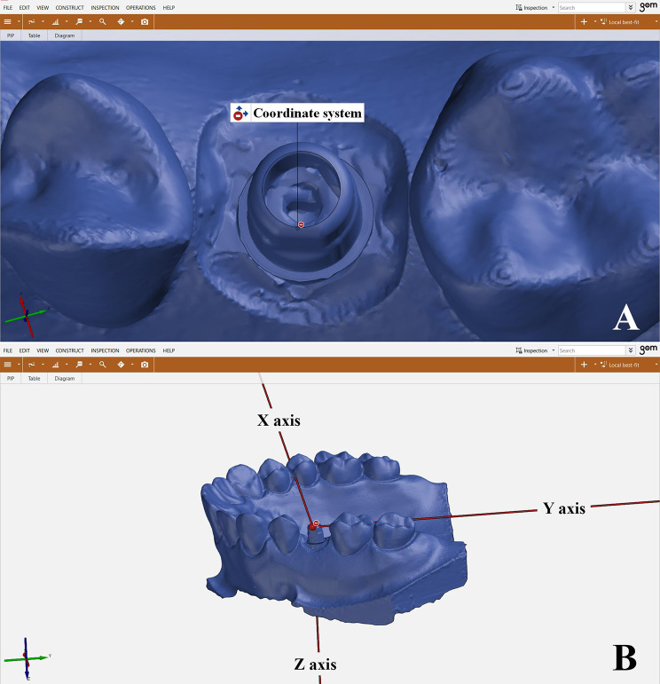
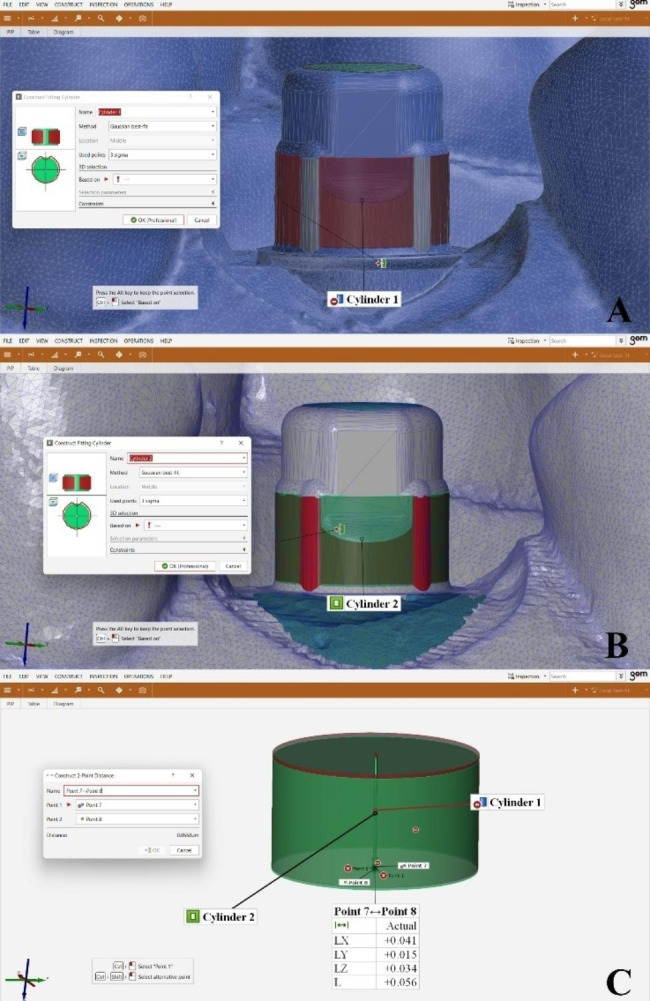
Methods: A maxillary model with a dummy implant in the 2nd premolar (Proactive Tapered Implant; Neoss) was 3D printed and TB (Ti Neolink Mono; Neoss) was tightened on the implant and scanned by using a laboratory scanner (inEos X5; Dentsply Sirona) (reference scan). An SB (Elos Medtech) and an HA-SP (Neoss) were subsequently connected to the implant and were scanned 10 times each by using the same scanner (test scans). All the scans were exported as STL files and imported into CAD software where the TBs were formed. Test scans were superimposed on reference scans for transfer accuracy analysis using 3D metrology software (GOM Inspect; GOM GmbH) in terms of angular deviation in vertical and horizontal directions, linear deviation in each XYZ axis of TBs and total linear deviation in all axes. Statistical analysis was done using independent sample t test. When Levene's test for equality of variances was significant, Welch's t-test was used. (P value < 0.05) RESULTS: Significant differences were found amongst the tested groups in both angular and linear deviation in terms of trueness with less deviation values for the SB group (P < 0.001). For the precision, significant differences were found amongst the tested groups in angular deviation in vertical direction with less deviation value for the SB group compared to HA-SP group (P < 0.001). However, no significant difference was found between the tested groups regarding the angular deviation in horizontal direction (P = 1.000). Moreover, significant differences were found amongst the tested groups in linear deviations with less linear deviations in XYZ axes for SB compared to HA-SP group (P = 0.020, < 0.001, = 0.010 respectively).

Conclusions: SB showed less angular and linear deviation values in the 3D positional transfer of TB than HA-SP indicating higher degree of accuracy of SB.

Keywords: Healing abutment-scan peg; Meteorology software; Positional transfer accuracy; Scan body; Titanium base abutment.


Conflict of interest statement

The authors declare no competing interests.

Figures

Fig. 1
Fig. 1
Investigated scan bodies. (A) One-piece SB, (B) Two-piece HA-SP.
Fig. 2
Fig. 2
Design of maxillary model with an implant site in 2nd premolar, (A) Occlusal view, (B) Lateral view
Fig. 3
Fig. 3
A. 3D printed implant model, B. The vertical mark on the screwdriver was positioned mid-buccally during implant placement
Fig. 4
Fig. 4
Data acqusition (A) TB abutment tightened on the implant so that the flat surface is positioned buccally, (B) Desktop scanning of TB, (C) SB tightened on the implant, (D) Desktop scanning of model with SB, (E) HA-SP tightened on the implant, (F) Desktop scanning of model with HA-SP.
Fig. 5
Fig. 5
Steps to obtain the reference scan (A) STL-2 was imported to CAD software, (B) “Best-fit matching” between SB in STL-1 and SB in CAD library, (C) TB was formed and exported solely as STL-4, (D) STL-1 was imported to CAD software, (E) STL-1 and STL-4 were aligned together through fixing points, (F) Reference scan was formed with well-defined TB structure
Fig. 6
Fig. 6
Steps to obtain SB test scans (A) STL-2 was imported to CAD software, (B) Virtual alignment of SB using the “best-fit matching” tool, (C) TB position corresponding to SB was formed (SB test scan)
Fig. 7
Fig. 7
Steps to obtain HA-SP test scans (A) STL-3 was imported to CAD software, (B) Virtual alignment of HA-SP using the “best-fit matching” tool, (C) TB position corresponding to HA-SP was formed (HA-SP test scan)
Fig. 8
Fig. 8
Local best-fit alignment of test and reference scans on 3D meterology software by selecting all teeth except TB site. The green color on the teeth showed zero deviation between the aligned test and reference scans, while the TB site showed red to blue color refering to the deviation that need to be analyzed (A) SB and reference scan, (B) HA-SP and reference scan
Fig. 9
Fig. 9
A. A coordinate system originating from a selected point on the occlusobuccal surface of the TB was used, B. XYZ axes resembled buccopalatal, mesiodistal and occlusocervical axes respectively
Fig. 10
Fig. 10
Angular deviation analysis A. Points and lines created on reference scan, B. Points and lines created on test scans, C. Angles between line 1 and line 3 resembled angular deviation in vertical direction and angles between line 2 and line 4 resembled angular deviation in horizontal direction
Fig. 11
Fig. 11
Linear deviation analysis (A) Fitting cylinder created on reference scan, (B) Fitting cylinder created on test scans, (C) A 2-point distance was constructed between both fitting cylinders’ center points corresponding to linear deviations in XYZ axes

References

    1. Flügge T, van der Meer WJ, Gonzalez BG, Vach K, Wismeijer D, Wang P. The accuracy of different dental impression techniques for implant-supported dental prostheses: a systematic review and meta-analysis. Clin Oral Implants Res. 2018;29:374–92. doi: 10.1111/clr.13273. - DOI - PubMed
    1. Mizumoto RM, Yilmaz B. Intraoral scan bodies in implant dentistry: a systematic review. J Prosthet Dent. 2018;120:343–52. doi: 10.1016/j.prosdent.2017.10.029. - DOI - PubMed
    1. Rutkunas V, Geciauskaite A, Jegelevicius D, Vaitiekunas M. Accuracy of digital implant impressions with intraoral scanners. A systematic review. Eur J Oral Implantol. 2017;10:101–20. - PubMed
    1. Schmidt A, Wöstmann B, Schlenz MA. Accuracy of digital implant impressions in clinical studies: a systematic review. Clin Oral Implants Res. 2022;33:573–85. doi: 10.1111/clr.13951. - DOI - PubMed
    1. Schmidt A, Billig JW, Schlenz MA, Wöstmann B. The influence of using different types of scan bodies on the transfer accuracy of Implant position: an in Vitro Study. Int J Prosthodont. 2021;34:254–60. doi: 10.11607/ijp.6796. - DOI - PubMed

LinkOut - more resources