

Pseudoaveraging for denoising of OCT angiography: a deep learning approach for image quality enhancement in healthy and diabetic eyes
- PMID: 37822004
- PMCID: PMC10568842
- DOI: 10.1186/s40942-023-00486-5
Pseudoaveraging for denoising of OCT angiography: a deep learning approach for image quality enhancement in healthy and diabetic eyes
Abstract
Background: This study aimed to develop a deep learning (DL) algorithm that enhances the quality of a single-frame enface OCTA scan to make it comparable to 4-frame averaged scan without the need for the repeated acquisitions required for averaging.
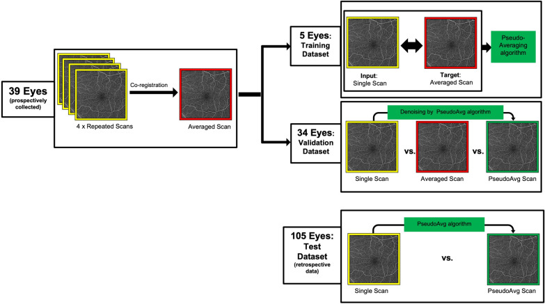
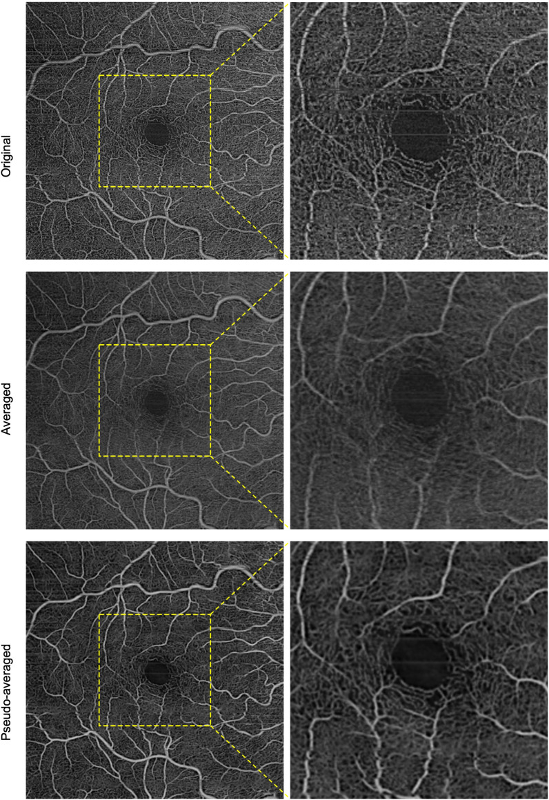
Methods: Each of the healthy eyes and eyes from diabetic subjects that were prospectively enrolled in this cross-sectional study underwent four repeated 6 × 6 mm macular scans (PLEX Elite 9000 SS-OCT), and the repeated scans of each eye were co-registered to produce 4-frame averages. This prospective dataset of original (single-frame) enface scans and their corresponding averaged scans was divided into a training dataset and a validation dataset. In the training dataset, a DL algorithm (named pseudoaveraging) was trained using original scans as input and 4-frame averages as target. In the validation dataset, the pseudoaveraging algorithm was applied to single-frame scans to produce pseudoaveraged scans, and the single-frame and its corresponding averaged and pseudoaveraged scans were all qualitatively compared. In a separate retrospectively collected dataset of single-frame scans from eyes of diabetic subjects, the DL algorithm was applied, and the produced pseudoaveraged scan was qualitatively compared against its corresponding original.
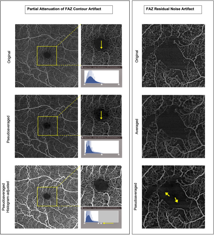
Results: This study included 39 eyes that comprised the prospective dataset (split into 5 eyes for training and 34 eyes for validating the DL algorithm), and 105 eyes that comprised the retrospective test dataset. Of the total 144 study eyes, 58% had any level of diabetic retinopathy (with and without diabetic macular edema), and the rest were from healthy eyes or eyes of diabetic subjects but without diabetic retinopathy and without macular edema. Grading results in the validation dataset showed that the pseudoaveraged enface scan ranked best in overall scan quality, background noise reduction, and visibility of microaneurysms (p < 0.05). Averaged scan ranked best for motion artifact reduction (p < 0.05). Grading results in the test dataset showed that pseudoaveraging resulted in enhanced small vessels, reduction of background noise, and motion artifact in 100%, 82%, and 98% of scans, respectively. Rates of false-positive/-negative perfusion were zero.
Conclusion: Pseudoaveraging is a feasible DL approach to more efficiently improve enface OCTA scan quality without introducing notable image artifacts.
Keywords: Averaging; Deep learning; Denoising; Diabetic retinopathy; Image artifact; Image quality; OCTA; Optical coherence tomography angiography; Pseudoaveraging.
© 2023. Brazilian Retina and Vitreous Society.
Conflict of interest statement
Warren Lewis MS is a consultant for Zeiss; Luis De Sisternes PhD is a former employee at Zeiss (employee at the time of the project); Stephanie Magazzeni PhD is an employee at Zeiss; Sophie Kubach MS is a former employee at Zeiss (employee at the time of the project); Mary Durbin PhD is a former employee (employee at the time of the project); AYA is an employee at Boston Image Reading Center; Caroline Baumal MD is a consultant for Genentech, Regeneron, Roche, Apellis, and Eyepoint. Jay S. Duker MD is an employee at EyePoint Pharmaceuticals, a consultant for Aura Biosciences, and a board member of Sesen Bio and Hubble Therapeutics; Nadia K. Waheed MD MPH is a consultant for Topcon, Complement Therapeutics, Olix Pharma, Iolyx Pharmaceuticals, Hubble, Saliogen, and Syncona, has received speaker fees from Nidek and Zeiss and equity interest from Ocudyne, Gyroscope, an employee at AGTC, has received research support to institution from Zeiss, Topcon, and Nidek (all active, all devices still at institution as part of research).
The other authors have no competing interests.
Figures
References
-
- Spaide RF, James GF, Nadia KW, Srinivas R, Sadda A, Staurenghi G. Optical coherence tomography angiography. HHS Public Access. 2019 doi: 10.1016/j.preteyeres.2017.11.003.Optical. - DOI
Grants and funding
LinkOut - more resources
Full Text Sources
