. 2023 Oct 6:18:5651-5670.
doi: 10.2147/IJN.S426849. eCollection 2023.

The Therapeutic Effects of MUC1-C shRNA@Fe3O4 Magnetic Nanoparticles in Alternating Magnetic Fields on Triple-Negative Breast Cancer

Affiliations

The Therapeutic Effects of MUC1-C shRNA@Fe3O4 Magnetic Nanoparticles in Alternating Magnetic Fields on Triple-Negative Breast Cancer

Zhifeng Li et al. Int J Nanomedicine. .

Abstract

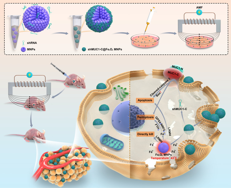
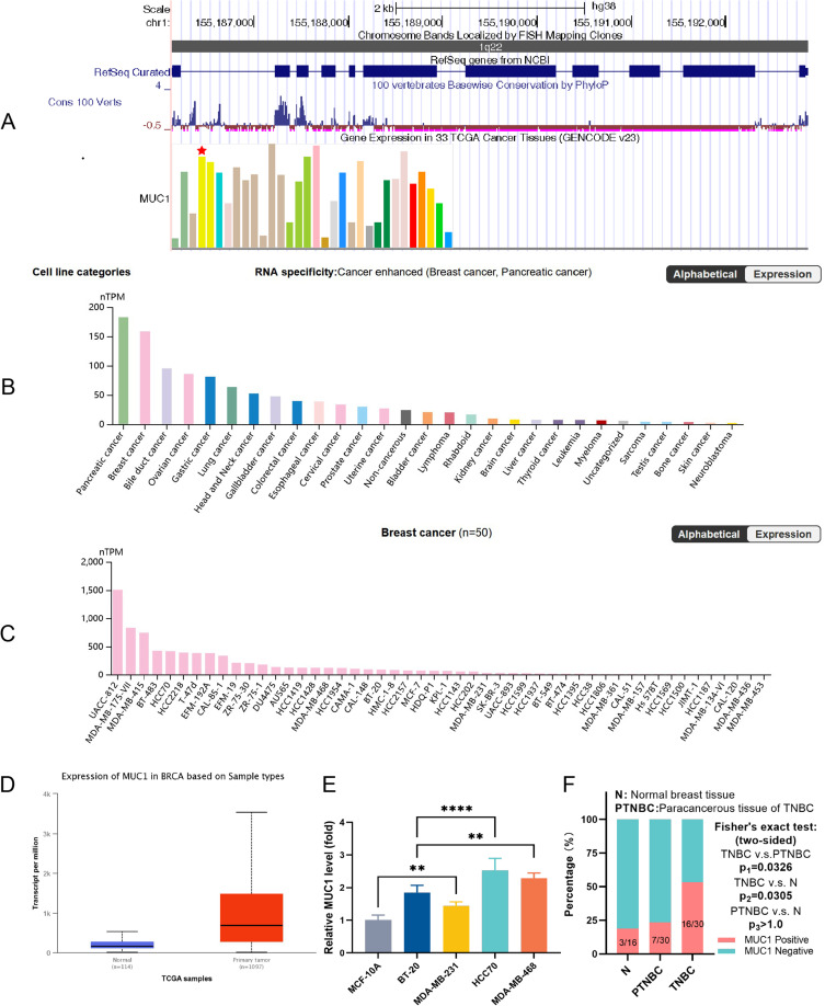
Purpose: Improving the treatment of triple-negative breast cancer (TNBC) is a serious challenge today. The primary objective of this study was to construct MUC1-C shRNA@ Fe3O4 magnetic nanoparticles (MNPs) and investigate their potential therapeutic benefits in alternating magnetic fields (AMF) on TNBC.

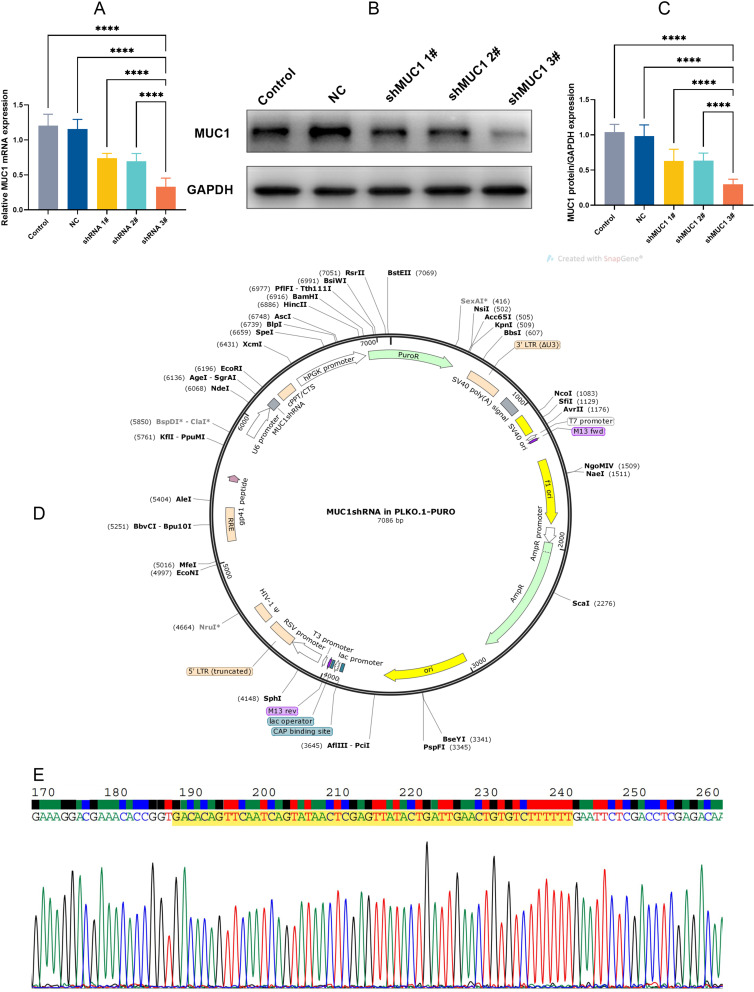
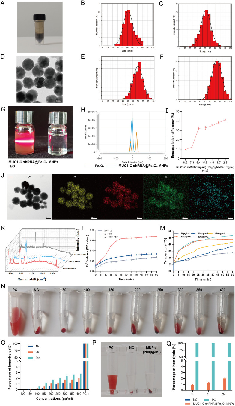
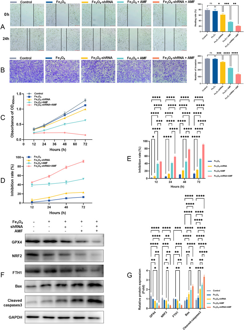
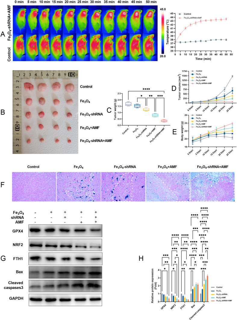
Methods: Firstly, we verified the high expression of MUC1 in TNBC and synthesized specific MUC1-C shRNA plasmids (MUC1-C shRNA). Then, we prepared and characterized MUC1-C shRNA@Fe3O4 MNPs and confirmed their MUC1-C gene silencing effect and magneto-thermal conversion ability in AMF. Moreover, the inhibitory effects on TNBC in vitro and in vivo were observed as well as biosafety. Finally, the protein levels of BCL-2-associated X protein (Bax), cleaved-caspase3, glutathione peroxidase inhibitor 4 (GPX4), nuclear factor erythroid 2-related factor 2 (NRF2), and ferritin heavy chain 1 (FTH1) in TNBC cells and tissues were examined, and it was speculated that apoptosis and ferroptosis were involved in the synergistic treatment.

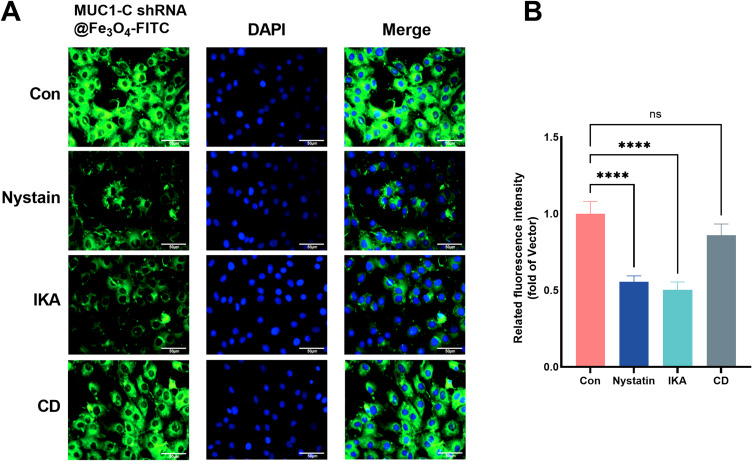
Results: MUC1-C shRNA@ Fe3O4 MNPs have a size of ~75 nm, with an encapsulation rate of (29.78±0.63) %, showing excellent gene therapy and magnetic hyperthermia functions. Under a constant AMF (3Kw) and a set concentration (200µg mL-1), the nanoparticles could be rapidly warmed up within 20 minutes and stabilized at about 43 °C. It could be uptaken by TNBC cells through endocytosis and significantly inhibit their proliferation and migration, with a growth inhibition rate of 79.22% for TNBC tumors. After treatment, GPX4, NRF2, and FTH1 expression levels in TNBC cells and tumor tissues were suppressed, while Bax and cleaved-caspase3 were increased. As key therapeutic measures, gene therapy, and magnetic hyperthermia have shown a synergistic effect in this treatment strategy, with a combined index (q index) of 1.23.

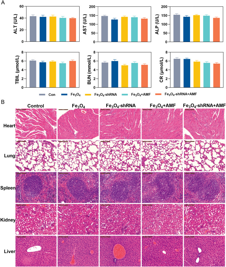
Conclusion: In conclusion, we developed MUC1-C shRNA@Fe3O4 MNPs with magnetic hyperthermia and gene therapy functions, which have shown satisfactory therapeutic effects on TNBC without significant side effects. This study provides a potential option for the precision treatment of TNBC.

Keywords: MUC1; ferroptosis; hyperthermia; nanoparticle; triple-negative breast cancer.


Conflict of interest statement

The authors declare no conflicts of interest in this work.

Figures

Scheme 1
Scheme 1
Signal pathway and mechanism of MUC1-C regulating TNBC Occurrence and progression (Comprehensive arrangement according to the published articles).
Scheme 2
Scheme 2
Schematic illustration of MUC1-C shRNA@Fe3O4 MNPs construction and the therapeutic mechanism on TNBC. MUC1-C shRNA and Fe3O4 MNPs were self-assembled into MUC1-C shRNA@ Fe3O4 MNPs by electrostatic adsorption. After entering TNBC cells and under the action of AMF, on the one hand, it induces apoptosis and ferroptosis by targeting MUC1-C, on the other hand, it increases the temperature (43 °C) and the Fe2+ concentration directly kills TNBC cells and stimulates apoptosis and ferroptosis mechanisms.
Figure 1
Figure 1
MUC1 and its expressions in various types of tumors (from UCSC genome browser on human, the red pentagram indicates breast invasive carcinoma) (A), MUC1 protein expression in different tumor cell lines (B) and BRCA cell lines (C) (based on datasets of Human Protein Atlas), MUC1 expression between BRCA and normal tissue (D) (based on datasets of The Cancer Genome Atlas), MUC1 expression in normal human breast cell (MCF-10A) and TNBC cell lines (BT-20, MDA-MB-231, MDA-MB-468, HCC-70) (E). IHC results of MUC1 in TNBC tissues, paraneoplastic tissues and normal breast tissues (F). Error bars represent means ± SD. **p<0.01, ****p<0.001.
Figure 2
Figure 2
Silence efficiency of MUC1-C shRNA on TNBC cell line HCC-70. RT-qPCR (A) and Western blot detected the silencing efficiency of MUC1-C shRNA (B and C). MUC1-C shRNA (3#) plasmid map (D) and target sequence (the highlighted yellow section) (E). Error bars represent means ± SD. ****p<0.001.
Figure 3
Figure 3
Characterization of MUC1-C shRNA@Fe3O4 MNPs. Appearance (A) and TEM image (D) of the prepared MUC1-C shRNA@Fe3O4 MNPs. DSL Size distribution of Fe3O4 MNPs (B and C) and MUC1-C shRNA@Fe3O4 MNPs (E and F). Tyndall effect of MUC1-C shRNA@Fe3O4 MNPs (G). Zeta potential of MUC1-C shRNA@Fe3O4 MNPs (H). Encapsulation efficiency at different shRNA/MNPs ratios (I). TEM elemental mapping analysis of MUC1-C shRNA@Fe3O4 MNPs (J). Raman spectrum of MUC1-C shRNA, Fe3O4 MNPs, and MUC1-C shRNA@Fe3O4 MNPs (K). Release profiles of Fe2+ from MUC1-C shRNA@Fe3O4 MNPs at different pH values with or without AMF (L). The heating curves of MUC1-C shRNA@Fe3O4 MNPs with different concentrations (50–250μg/mL) (M). Hemolysis test at different concentrations (MUC1-C shRNA@Fe3O4 MNPs) (n=3) (N and O). Hemolysis test (200μg/mL, n=3) (P and Q). Error bars represent means ± SD. TEM scale bar: 50nm.
Figure 4
Figure 4
Cellular uptake of NPs. Cellular uptake of MUC1-C shRNA@Fe3O4 MNPs in the HCC-70 cells was observed under a CLSM. Green fluorescence indicates MUC1-C shRNA@Fe3O4-FITC and blue fluorescence indicates the nucleus (A). Investigation of internalization pathway of MUC1-C shRNA@Fe3O4 MNPs in HCC-70 cells in the presence of different endocytosis inhibitors (n=3) (B). Error bars represent means ± SD. Scale bar: 50μm. ****p<0.001.
Figure 5
Figure 5
MUC1-C shRNA@Fe3O4 MNPs suppress the migration, invasion, and proliferation in HCC-70 cell lines. Wound healing (A), Transwell (B), CCK-8 OD450nm (C), and inhibition rate based on CCK-8 (D and E). The protein bands (F) and quantitative statistical results (G) of Bax, cleaved-caspase3, GPX4, NRF2, and FTH1 of HCC-70 cell lines (n=3). Error bars represent means ± SD. *p<0.05, **p<0.01, ***p<0.005, ****p<0.001.
Figure 6
Figure 6
The in vivo therapeutic effects of the MUC1-C shRNA@Fe3O4 MNPs on TNBC. IR thermal images of TNBC-bearing C-NKG mice with local injection of MUC1-C shRNA@Fe3O4 MNPs (200 µg mL−1), or normal saline under AMF (3Kw) (A). Images (B) and weights (C) of xenograft tumors harvested from the mice after various treatments on the 25th day. Tumor volumes (D) and Body weight (E) growth in mice were measured after different treatments every 5 days within 25 days (n=4). Prussian blue reaction of tumor tissue showing iron staining and tumor necrosis, labeled area was typical necrosis (200x) (F). Expression of Bax, cleaved-caspase 3, GPX4, NRF2, and FTH1 in tumor tissues (G). Quantitative statistical results according to the protein bands (H). Error bars represent means ± SD. *p<0.05, **p<0.01, ***p<0.005, ****p<0.001.
Figure 7
Figure 7
Serum biochemistry indicators assessment of TNBC mice (n=4). Alanine aminotransferase (ALT), Aspartate aminotransferase (AST), Alkaline phosphatase (ALP), Total bilirubin (TBIL). Blood urea nitrogen (BUN), Creatinine (CR) (A). Effect of MUC1-C shRNA@Fe3O4 MNPs on the histopathology of heart, lung, spleen, Kidney, and liver from TNBC mice (B). Error bars represent means ± SD. Scale bar: 100μm.

References

    1. Siegel RL, Miller KD, Fuchs HE, Jemal A. Cancer statistics, 2022. CA Cancer J Clin. 2022;72(1):7–33. doi:10.3322/caac.21708 - DOI - PubMed
    1. Howard FM, Olopade OI. Epidemiology of triple-negative breast cancer: a review. Cancer J. 2021;27(1):8–16. doi:10.1097/PPO.0000000000000500 - DOI - PubMed
    1. Rugo HS, Olopade OI, DeMichele A, et al. Adaptive randomization of veliparib-carboplatin treatment in breast cancer. N Engl J Med. 2016;375(1):23–34. doi:10.1056/NEJMoa1513749 - DOI - PMC - PubMed
    1. Schmid P, Cortes J, Dent R, et al. Event-free survival with pembrolizumab in early triple-negative breast cancer. N Engl J Med. 2022;386(6):556–567. doi:10.1056/NEJMoa2112651 - DOI - PubMed
    1. Downs-Canner S, Mittendorf EA. Preoperative immunotherapy combined with chemotherapy for triple-negative breast cancer: perspective on the KEYNOTE-522 study. Ann Surg Oncol. 2023;30(6):3166–3169. doi:10.1245/s10434-023-13267-z - DOI - PubMed

LinkOut - more resources