. 2023 Sep 29:35:101550.
doi: 10.1016/j.bbrep.2023.101550. eCollection 2023 Sep.

Activated protein C signaling mediates neuroinflammation in seizure induced by pilocarpine

Affiliations

Activated protein C signaling mediates neuroinflammation in seizure induced by pilocarpine

Linda Ines Zoungrana et al. Biochem Biophys Rep. .

Abstract

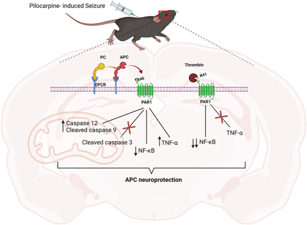
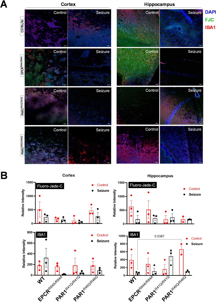
Epilepsy is one of the most common and oldest neurological disorders, characterized by periodic seizures that affect millions globally. Despite its long history, its pathophysiology is not fully understood. Additionally, the current treatment methods have their limitations. Finding a new alternative is necessary. Activated Protein C (APC) has been proven to have neurological protection in other neurological disorders; however, there is no study that focuses on the role of APC in seizures. We propose that APC's protective effect could be associated with seizures through inflammation and apoptosis regulation. The results demonstrated that APC's pathway proteins are involved in neuroprotection mechanisms in seizure-induced models by acting on certain inflammatory factors, such as NF-κB and apoptosis proteins.

Keywords: Activated protein C; Epilepsy; Inflammation.


Conflict of interest statement

The authors declare no conflict of interest.

Figures

Image 1
Graphical abstract
Fig. 1
Fig. 1
Behavioral analysis in an Open Field Test. (A) Illustrative representation of travel pathway (purple) of WT C57BL/6J, EPCRR84A/R84A, PAR1R41Q/R41Q, PAR1R46Q/R46Q of 15 min recording in the open field test arena. The center is indicated by the smaller square and the larger square represents the perimeter. WT C57BL/6J travel a significantly longer distance. (B) Compared to EPCRR84A/R84A, PAR1R46Q/R46Q. The difference in the time spent on the perimeter or the center are not significant. Histograms bars indicate the mean ± SEM; One way ANOVA, post hoc analysis by Tukey's HSD (WT C57BL/6J: n = 16, EPCRR84A/R84A: n = 21, PAR1R41Q/R41Q: n = 20, PAR1R46Q/R46Q: n = 19). (For interpretation of the references to colour in this figure legend, the reader is referred to the Web version of this article.)
Fig. 2
Fig. 2
EEG Recording and Seizure Severity. (A) Illustration of EEG electrode implantation in the mice's upper felt and lower right of the scalp. (B) Representative EEG recording of WT C57BL/6J, EPCRR84A/R84A, PAR1R41Q/R41Q, PAR1R46Q/R46Q baseline and during scopolamine, pilocarpine and midazolam injection (i.p). (C) Significant increase in the severity of the seizure compared to baseline but the difference in the power band, spikes number and spikes train between WT C57BL/6J, EPCRR84A/R84A, PAR1R41Q/R41Q, PAR1R46Q/R46Q are not significant. Data provided by DSI Harvard Bioscience NeuroScore software. Histograms bars indicate the mean ± SEM; Two-way ANOVA, post hoc analysis by Tukey's HSD for power band and Racine score, One-way ANOVA for spikes numbers and spikes train.
Fig. 3
Fig. 3
Immunoblotting of inflammatory protein. Protein expression of inflammatory proteins p–NF–κB and NF-κB in the cortex (A) and hippocampus (B) region of the brain under control or seizure conditions of WT C57BL/6J, EPCRR84A/R84A, PAR1R41Q/R41Q, PAR1R46Q/R46Q.
Fig. 4
Fig. 4
Immunofluorescence of degenerative neuron and microglia activity. (A) Representative staining of DAPI (blue), FJC (green), and IBA1 (red) in the cortex and hippocampus under control or 72-h post-seizure of WT C57BL/6J, EPCRR84A/R84A, PAR1R41Q/R41Q, PAR1R46Q/R46Q. Scale bars are 50 μm. (B) Relative intensity of FJC and IBA1 comparing control, and 72-h post-seizure of WT C57BL/6J, EPCRR84A/R84A, PAR1R41Q/R41Q, PAR1R46Q/R46Q. Histogram bars indicate the mean ± SEM; Two-way ANOVA, post hoc analysis by Tukey's HSD. (For interpretation of the references to colour in this figure legend, the reader is referred to the Web version of this article.)

References

    1. Center for Disease Control and Prevention . 2023. Epilepsy.https://www.cdc.gov/epilepsy/index.html
    1. World Health Organization . 2023. Epilepsy.https://www.who.int/news-room/fact-sheets/detail/epilepsy
    1. Yuen A.W.C., Keezer M.R., Sander J.W. Epilepsy is a neurological and a systemic disorder. Epilepsy Behav. 2018;78:57–61. doi: 10.1016/j.yebeh.2017.10.010. - DOI - PubMed
    1. Kumar P., Lim A., Hazirah S.N., Chua C.J.H., Ngoh A., Poh S.L., Yeo T.H., Lim J., Ling S., Sutamam N.B., Petretto E., Low D.C.Y., Zeng L., Tan E.-K., Arkachaisri T., Yeo J.G., Ginhoux F., Chan D., Albani S. Single-cell transcriptomics and surface epitope detection in human brain epileptic lesions identifies pro-inflammatory signaling. Nat. Neurosci. 2022;25:956–966. doi: 10.1038/s41593-022-01095-5. - DOI - PMC - PubMed
    1. Akyuz E., Polat A.K., Eroglu E., Kullu I., Angelopoulou E., Paudel Y.N. Revisiting the role of neurotransmitters in epilepsy: an updated review. Life Sci. 2021;265 doi: 10.1016/j.lfs.2020.118826. - DOI - PubMed

LinkOut - more resources