. 2023 Aug 21;11(10):6560-6570.
doi: 10.1002/fsn3.3605. eCollection 2023 Oct.

Immunostimulatory effects of marine algae extracts on in vitro antigen-presenting cell activation and in vivo immune cell recruitment

Affiliations

Immunostimulatory effects of marine algae extracts on in vitro antigen-presenting cell activation and in vivo immune cell recruitment

Thi Len Ho et al. Food Sci Nutr. .

Abstract

Marine algae are photosynthetic eukaryotic organisms that are widely used as sources of food, cosmetics, and drugs. However, their biological and immunological effects on immune cells have not been fully elucidated. To unravel their immunological activity and broaden their application, we generated antigen-presenting cells (APCs), including dendritic cells (DCs) and macrophages, from mouse bone marrow cells and treated them with six different marine algae extracts (MAEs). We evaluated cell viability, activation marker expression, and pro-inflammatory cytokine production by APCs after 2 days of MAE treatment. All six MAEs significantly induced cytokine production of APCs, among which Pyropia yezoensis (PY), Peyssonnelia caulifera (PC), and Meristotheca papulosa (MP) extracts exhibited the strongest effect. Cladophora wrightiana var. minor (CW) extract moderately upregulated cytokine levels but increased the expression of activation markers on DCs. Moreover, PY, PC, MP, Sargassum pectinifera (SP), and Caulerpa okamurae (CO) pre-treated APCs effectively stimulated T-cell proliferation and cytokine production. Furthermore, the mice injected with MAEs exhibited higher cytokine (TNF-α, IL-6, and IL-1β) production as well as enhanced innate immune cell recruitment capacities (DCs, monocytes, neutrophils, and natural killer cells) in the peritoneal cavity of the mice compared to those of the non-treated mice. Therefore, all MAEs exhibited immunostimulatory potential, with PY, PC, CW, and MP extracts being the most effective in stimulating immune responses and cell activation. To the best of our knowledge, this is the first study to determine the immunomodulatory activities of six MAEs both in vitro and in vivo.

Keywords: DCs; immunostimulatory effects; macrophages; marine algae extracts.


Conflict of interest statement

The authors declare that there is no conflict of interest.

Figures

FIGURE 1
FIGURE 1
Cell viability after treatment with marine algae extracts. Bone marrow‐derived dendritic cells (a) and macrophages (b) were treated with different concentrations of each marine algae extract (marine algae extract; 4, 20, and 100 μg/mL) for 2 days, and MTT assay was performed. Data are shown as mean ± standard error.
FIGURE 2
FIGURE 2
Cytokine production of marine algae extract (MAE)‐treated bone marrow‐derived dendritic cells (BMDCs) and bone marrow‐derived macrophages (BMDMs). BMDCs (a) and BMDMs (b) were treated in vitro with MAEs or monophosphoryl lipid A (MPL) (positive control, 0.1 μg/mL MPL; 20 μg/mL each MAE). After 2 days, the level of nitric oxide (NO), interleukin (IL)‐6, tumor necrosis factor (TNF)‐alpha, and IL‐12p40 was determined by ELISA. Data are shown as mean ± standard error. One‐way ANOVA and Tukey's multiple comparison tests were performed for statistical analysis. *p < .0332; **p < .0021; ***p < .0002; and ****p < .0001 compared to control group.
FIGURE 3
FIGURE 3
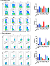
Expression of activation markers by bone marrow‐derived dendritic cells (BMDCs) and bone marrow‐derived macrophages (BMDMs) induced by marine algae extracts (MAEs). BMDCs and BMDMs were treated with MAEs or monophosphoryl lipid A (MPL) (positive control) in vitro (0.1 μg/mL MPL or 20 μg/mL each MAE). After 2 days, MHC class II, CD40, and CD86 expression on dendritic cells (DCs) (a) and macrophages (b) were determined by flow cytometry and analyzed by FlowJo. DCs and macrophages were gated by CD11c and CD11b surface markers, respectively. The representative data obtained from the flow cytometry analysis were shown in pseudo‐color smooth plots. Data are shown as mean ± standard error. One‐way ANOVA and Tukey's multiple comparison tests were performed for statistical analysis. *p < .0332; **p < .0021; ***p < .0002; and ****p < .0001 compared to control group.
FIGURE 4
FIGURE 4
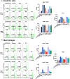
T‐cell proliferation and interferon gamma (IFN‐γ) production by T‐cells after co‐culture with marine algae extract (MAE)‐treated bone marrow‐derived dendritic cells (BMDCs) and bone marrow‐derived macrophages (BMDMs). BMDCs and BMDMs from BALB/c mice were pre‐treated with 0.1 μg/mL monophosphoryl lipid A (MPL) or 20 μg/mL of each MAE. After 2 days, the activated dendritic cells (a) and macrophages (b) were co‐cultured with CFSE‐labeled allogeneic naïve T‐cells, which were harvested from the spleen of C57BL/6 mice after 5 days. After co‐culture, the percentage of CFSE‐negative cells in either CD3+ CD4+ T‐cells or CD3+ CD8+ T‐cells was acquired by flow cytometry and analyzed by FlowJo, and cell supernatant was used to measure IFN‐γ secretion, using ELISA. The representative data obtained from the flow cytometry analysis were shown in histograms. Data are shown as mean ± standard error. One‐way ANOVA and Tukey's multiple comparison tests were performed for statistical analysis. *p < .0332; **p < .0021; ***p < .0002; and ****p < .0001 compared to control group.
FIGURE 5
FIGURE 5
Cytokine and chemokine levels after intraperitoneal injection of marine algae extracts (MAEs) in mice. BALB/c mice (n = 6) were intraperitoneally injected with monophosphoryl lipid A (MPL) (1 μg/mouse) or MAEs (100 μg/mouse). The levels of each cytokine and chemokine in the supernatant of the peritoneal exudates and peritoneal lavage were detected by ELISA after 6 h. Data are shown as mean ± standard error. One‐way ANOVA and Tukey's multiple comparison tests were performed for statistical analysis. *p < .0332 and **p < .0021 compared to control group.
FIGURE 6
FIGURE 6
Cell population in the peritoneal cavity of mice after injection with marine algae extracts (MAEs). BALB/c mice (n = 6) were intraperitoneally injected with monophosphoryl lipid A (MPL) (1 μg/mouse) or MAEs (100 μg/mouse). Peritoneal cells were collected from peritoneal lavage after 24 h of MAE injection via centrifugation. The percentage of dendritic cells, macrophages, monocytes, neutrophils, and natural killer cells in the peritoneal cavity was measured by flow cytometry and analyzed by FlowJo. Data are shown as mean ± standard error. One‐way ANOVA and Tukey's multiple comparison tests were performed for statistical analysis. *p < .0332; **p < .0021 compared to control group.

References

    1. Alves, A. , Sousa, E. , Kijjoa, A. , & Pinto, M. (2020). Marine‐derived compounds with potential use as cosmeceuticals and nutricosmetics. Molecules, 25(11), 2536. 10.3390/molecules25112536 - DOI - PMC - PubMed
    1. Andrew, M. , & Jayaraman, G. (2021). Marine sulfated polysaccharides as potential antiviral drug candidates to treat Corona Virus disease (COVID‐19). Carbohydrate Research, 505, 108326. - PMC - PubMed
    1. Barzkar, N. , Khan, Z. , Jahromi, S. T. , Pourmozaffar, S. , Gozari, M. , & Nahavandi, R. (2021). A critical review on marine serine protease and its inhibitors: A new wave of drugs? International Journal of Biological Macromolecules, 170, 674–687. - PubMed
    1. Bhardwaj, M. , Mani, S. , Malarvizhi, R. , Sali, V. K. , & Vasanthi, H. R. (2021). Immunomodulatory activity of brown algae Turbinaria ornata derived sulfated polysaccharide on LPS induced systemic inflammation. Phytomedicine, 89, 153615. - PubMed
    1. Chaperot, L. , Chokri, M. , Jacob, M. , Drillat, P. , Garban, F. , Egelhofer, H. , Molens, J. P. , Sotto, J. J. , Bensa, J. C. , & Plumas, J. (2000). Differentiation of antigen‐presenting cells (dendritic cells and macrophages) for therapeutic application in patients with lymphoma. Leukemia, 14(9), 1667–1677. - PubMed

LinkOut - more resources