. 2023 Oct 13;382(6667):eadf9941.
doi: 10.1126/science.adf9941. Epub 2023 Oct 13.

Spatiotemporal molecular dynamics of the developing human thalamus

Affiliations

Spatiotemporal molecular dynamics of the developing human thalamus

Chang N Kim et al. Science. .

Abstract

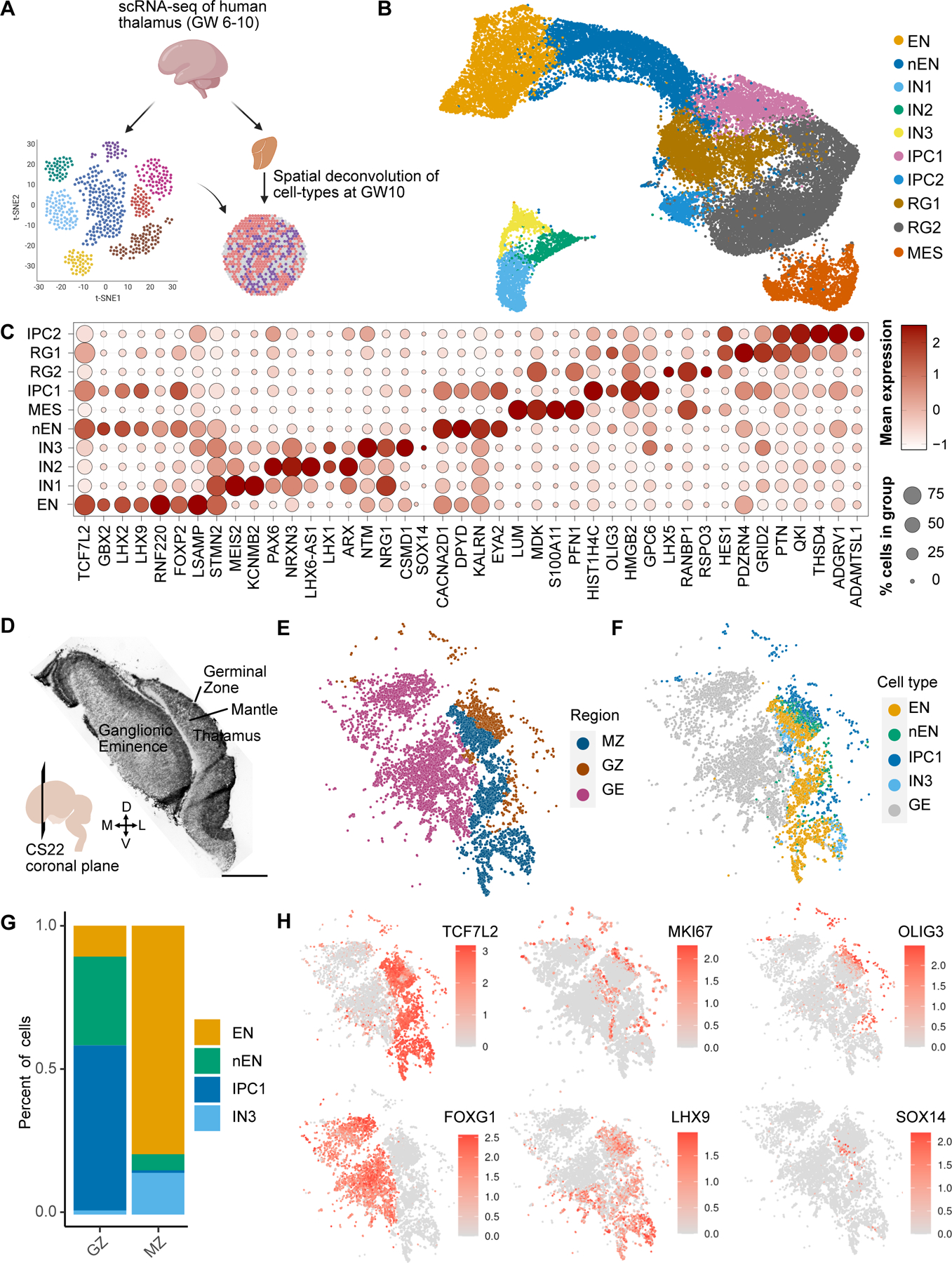
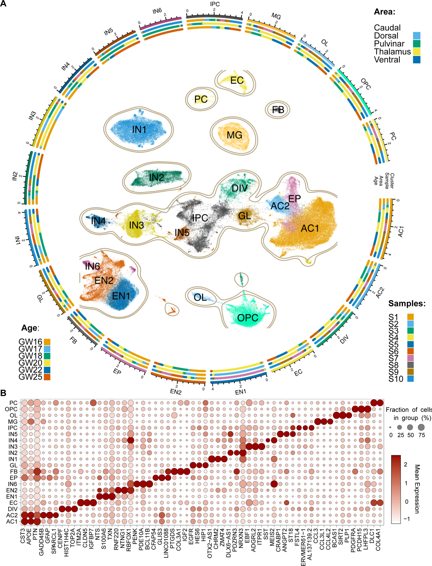
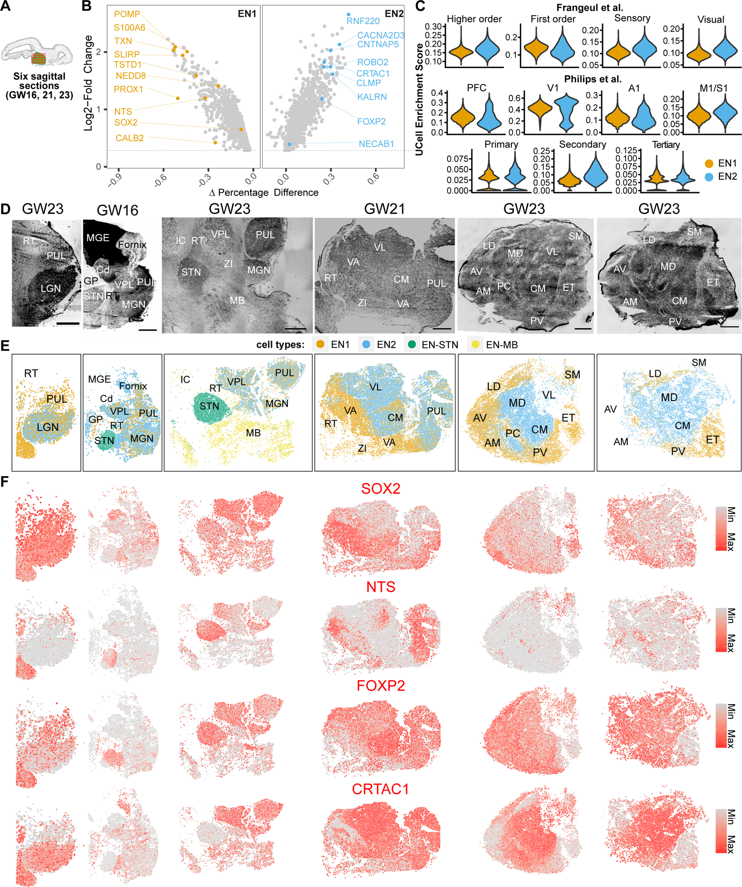
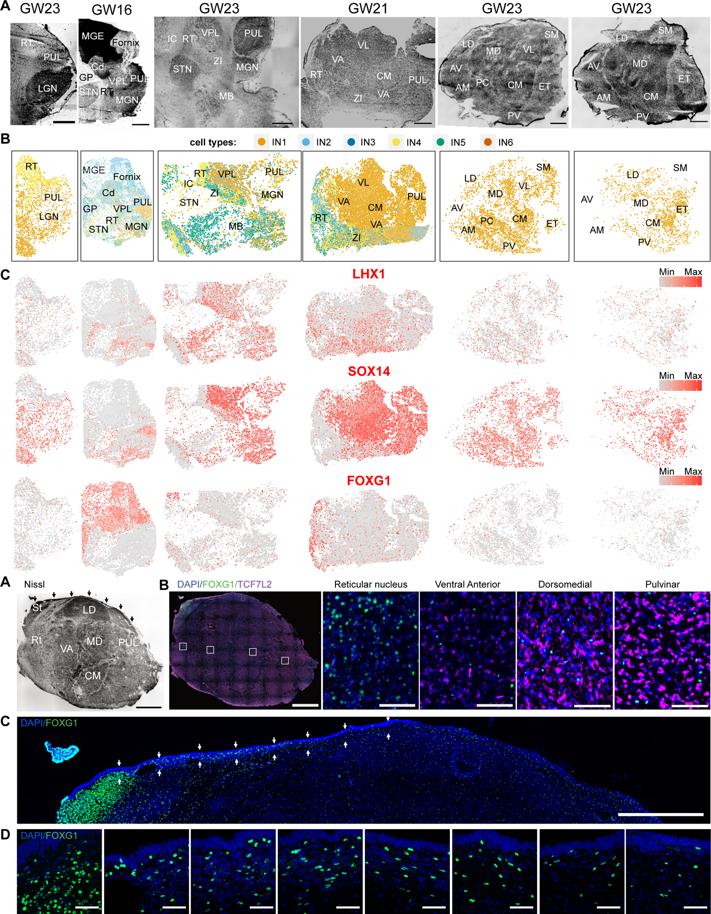
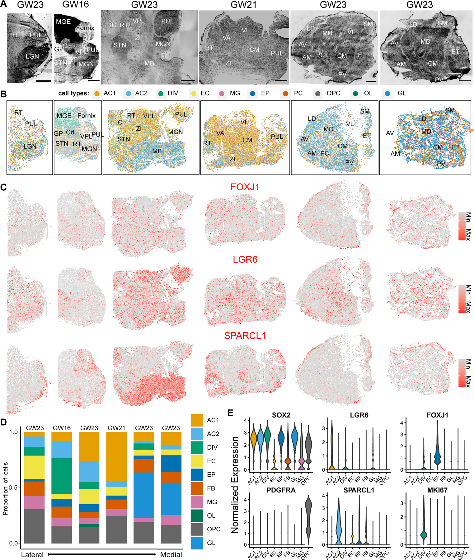
The thalamus plays a central coordinating role in the brain. Thalamic neurons are organized into spatially distinct nuclei, but the molecular architecture of thalamic development is poorly understood, especially in humans. To begin to delineate the molecular trajectories of cell fate specification and organization in the developing human thalamus, we used single-cell and multiplexed spatial transcriptomics. We show that molecularly defined thalamic neurons differentiate in the second trimester of human development and that these neurons organize into spatially and molecularly distinct nuclei. We identified major subtypes of glutamatergic neuron subtypes that are differentially enriched in anatomically distinct nuclei and six subtypes of γ-aminobutyric acid-mediated (GABAergic) neurons that are shared and distinct across thalamic nuclei.


Conflict of interest statement

Competing interests: Authors declare that they have no competing interests.

Figures

Fig. 1.
Fig. 1.. Neurogenesis in the first trimester thalamus.
A. Schematic overview of figure 1. We analyzed scRNA-seq data collected from three biologically independent first trimester thalamus samples (GW6-10). Spatial transcriptomics using MERFISH was performed to spatially map cell types identified by scRNA-seq. B. UMAP embedding of cell type annotations on the Harmony batch corrected space: EN - excitatory neuron, nEN – newborn excitatory neuron, IN1/2/3 - inhibitory neuron 1/2/3, RG1/2 - radial glia 1/2, IPC1/2 - intermediate progenitor cells 1/2, and MES - mesenchyme. C. Dot plot of marker genes across cell types/states. D. H&E of a MERFISH adjacent coronal section of CS22 (GW10) E. MERFISH region annotations. MZ - Thalamic Mantle Zone, GZ - Thalamic Germinal Zone, GE - Ganglionic Eminences. F. Spatially projected cell type annotations. G. Relative proportion of cell types within regions from E. H. Spatially projected expression of select marker genes, including telencephalic (FOXG1) and diencephalic (TCF7L2, LHX9) identity, dividing cells (MKI67), radial glia and IPCs (OLIG3), and rostral thalamus-derived GABAergic neurons (SOX14).
Fig. 2.
Fig. 2.. Cellular diversity in the second trimester thalamus.
A. Constellation plot representing cellular diversity in second trimester human thalamus. PC - pericytes, OPC - oligodendrocyte precursor cells, OL - oligodendrocytes, MG - microglia, IPC - intermediate progenitor cells, IN1-6 - Inhibitory GABAergic neurons 1–6, GL - glial progenitor cells, FB - fibroblasts, EP - ependymal cells, EN1-2 - excitatory glutamatergic neurons 1–2, EC - endothelial cells, DIV - dividing cells, AC1-2 - astrocytes 1–2. B. Dot plot represents enrichment of select marker gene expression across cell types in the second trimester human thalamus.
Fig. 3.
Fig. 3.. Spatially mapped thalamic glutamatergic neuron subtypes in human mid-gestation thalamus.
A. Eight sagittal sections total were collected from three biologically independent second trimester thalamus specimens (GW16, 21, 23). Six representative sections are shown here and the other two are shown in figs. S6 and S7. B. Marker genes enriched for glutamatergic subtypes EN1 and EN2. C. UCell enrichment scores for gene signatures relating to known classes of thalamus neurons from adult mouse studies calculated for glutamatergic neuron subtypes EN1 and EN2. PFC - prefrontal cortex, V1 - Visual cortex, A1 - Auditory cortex, M1/S1 Motor/Somatosensory cortex. D. Nissl stains of sagittal sections. Sections are in order from left (lateral- most) to right (medial-most). Scale bar: 2 mm. RT - Reticular Nucleus, PUL - Pulvinar, LGN - Lateral Geniculate Nucleus, MGE - Medial Ganglionic Eminence, Cd - Caudate, GP - Globus Pallidus, STN - Subthalamic Nucleus, VPL - Ventral Posterior Lateral Nucleus, MGN - Medial Geniculate Nucleus, MB - Midbrain, VA - Ventral Anterior Nucleus, VL - Ventral Lateral Nucleus, CM - Centromedian Nucleus, ZI - Zona Incerta, AV - Anteroventral Nucleus, AM - Anteromedial Nucleus, LD - Dorsolateral Nucleus, MD - Dorsomedial Nucleus, PC - Paracentral Nucleus, PV - Paraventricular Nucleus, SM - Stria Medullaris, ET - Epithalamus. E. Spatial clustering analysis of MERFISH transcriptomics data. EN1 and EN2 from spatial clustering analysis relate to EN1 and EN2 from scRNA-seq data based on gene expression profiles. EN-STN and EN-MB are distinct clusters not represented by our scRNA-seq, and relate to glutamatergic neurons in the subthalamic nucleus and midbrain, respectively. F. Spatial feature plots showing the expression of two example marker genes for EN1 (SOX2, NTS) and EN2 (FOXP2, CRTAC1) glutamatergic neuron subtypes in MERFISH datasets.
Fig. 4.
Fig. 4.. Spatially mapped GABAergic neuron subtypes across the human mid-gestation thalamus.
A. Nissl stains of sagittal sections. Sections are in order from left (lateral- most) to right (medial-most). Scale bar: 2 mm. RT - Reticular Nucleus, PUL - Pulvinar, LGN - Lateral Geniculate Nucleus, MGE - Medial Ganglionic Eminence, Cd - Caudate, GP - Globus Pallidus, STN - Subthalamic Nucleus, VPL - Ventral Posterior Lateral Nucleus, MGN - Medial Geniculate Nucleus, MB - Midbrain, VA - Ventral Anterior Nucleus, VL - Ventral Lateral Nucleus, CM - Centromedian Nucleus, ZI - Zona Incerta, AV - Anteroventral Nucleus, AM - Anteromedial Nucleus, LD - Dorsolateral Nucleus, MD - Dorsomedial Nucleus, PC - Paracentral Nucleus, PV - Paraventricular Nucleus, SM - Stria Medullaris, ET - Epithalamus. B. Spatial clustering analysis of MERFISH transcriptomics data depicting GABAergic neuron subtypes. C. Expression of GABAergic neuron subtype markers across the sections. D-E. Nissl (D) and Immunostaining (E) of FOXG1 and TCF7L2 on a lateral section in a sagittal section from a biologically independent GW19 thalamus. Arrows in Nissl stain highlight the corpus gangliothalamicus, which is a thin migratory stream from MGE to thalamus. Small dotted boxes are zoom-in images depicted on the right. Large dotted box is a zoom-in image depicted in Fig. 4F. Scale bar for Nissl and low magnification images: 2 mm. Scale bar for high magnification images: 50 µm. St: Stria terminalis. F. Zoom in of the dorsal region of the thalamus section depicted in (E) to highlight FOXG1 staining within and below the corpus gangliothalamicus (highlighted by the arrows). Scale bar: 2 mm. G. High magnification (63X) images of regions highlighted by arrows in Fig. 4F highlights FOXG1 staining within the corpus gangliothalamicus, in order from left to right. Scale bar: 50 µm.
Fig 5.
Fig 5.. Spatially mapped non-neural cell subtypes across the human mid-gestation thalamus.
A. Nissl stains of sagittal sections. Sections are in order from left (lateral-most) to right (medial-most). Scale bar: 2 mm. RT - Reticular Nucleus, PUL - Pulvinar, LGN - Lateral Geniculate Nucleus, MGE - Medial Ganglionic Eminence, Cd - Caudate, GP - Globus Pallidus, STN - Subthalamic Nucleus, VPL - Ventral Posterior Lateral Nucleus, MGN - Medial Geniculate Nucleus, MB - Midbrain, VA - Ventral Anterior Nucleus, VL - Ventral Lateral Nucleus, CM - Centromedian Nucleus, ZI - Zona Incerta, AV - Anteroventral Nucleus, AM - Anteromedial Nucleus, LD - Dorsolateral Nucleus, MD - Dorsomedial Nucleus, PC - Paracentral Nucleus, PV - Paraventricular Nucleus, SM - Stria Medullaris, ET - Epithalamus. B. Spatial clustering analysis of MERFISH transcriptomics data depicting non-neuronal subtypes. AC1 and AC2 - astrocyte subtypes, DIV - dividing cells, EC - endothelial cells, EP - ependymal cells, FB - fibroblasts, MG - microglia, OL - oligodendrocytes, OPC - oligodendrocyte progenitor cells, GL - bipotent glial progenitor cells. C. Spatial feature plots depicting normalized expression of EP (FOXJ1), AC1 (LGR6), and AC2 (SPARCL1) markers. D. Relative proportion of non-neural cell types across lateral-to-medial sections. E. Violin plots depicting normalized expression of glial, astrocytes, ependymal, and oligodendrocyte precursor marker genes across the 2 lateral-most GW23 sections.

Update of

References

    1. Guillery RW, Anatomical evidence concerning the role of the thalamus in corticocortical communication: a brief review. J Anat 187 ( Pt 3), 583–592 (1995). - PMC - PubMed
    1. Baldwin MKL, Balaram P, Kaas JH, The evolution and functions of nuclei of the visual pulvinar in primates. J Comp Neurol 525, 3207–3226 (2017). - PubMed
    1. Homman-Ludiye J, Bourne JA, The medial pulvinar: function, origin and association with neurodevelopmental disorders. J Anat 235, 507–520 (2019). - PMC - PubMed
    1. Molnar Z, Development and evolution of thalamocortical interactions. Eur J Morphol 38, 313–320 (2000). - PubMed
    1. Govek KW et al. Developmental trajectories of thalamic progenitors revealed by single-cell transcriptome profiling and Shh perturbation. Cell Rep 41, 111768 (2022). - PMC - PubMed