. 2023 Dec;12(32):e2301527.
doi: 10.1002/adhm.202301527. Epub 2023 Nov 3.

Noninvasive Treatment of Alzheimer's Disease with Scintillating Nanotubes

Affiliations

Noninvasive Treatment of Alzheimer's Disease with Scintillating Nanotubes

Sudipta Senapati et al. Adv Healthc Mater. 2023 Dec.

Abstract

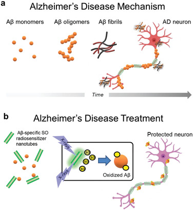
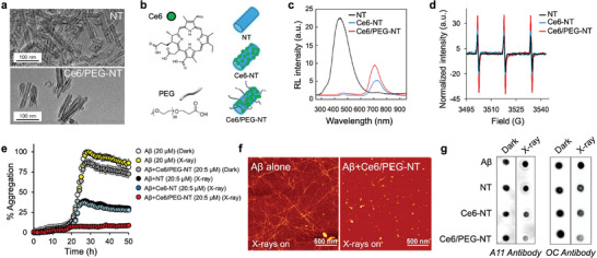
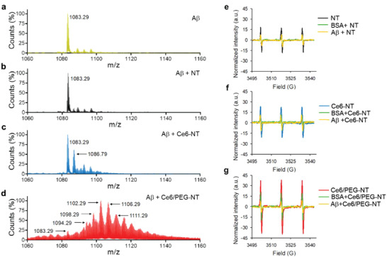
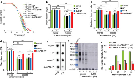
Effective and accessible treatments for Alzheimer's disease (AD) are urgently needed. Soluble Aβ oligomers are identified as neurotoxic species in AD and targeted in antibody-based drug development to mitigate cognitive decline. However, controversy exists concerning their efficacy and safety. In this study, an alternative strategy is proposed to inhibit the formation of Aβ oligomers by selectively oxidizing specific amino acids in the Aβ sequence, thereby preventing its aggregation. Targeted oxidation is achieved using biocompatible and blood-brain barrier-permeable multicomponent nanoscintillators that generate singlet oxygen upon X-ray interaction. Surface-modified scintillators interact selectively with Aβ and, upon X-ray irradiation, inhibit the formation of neurotoxic aggregates both in vitro and in vivo. Feeding transgenic Caenorhabditis elegans expressing human Aβ with the nanoscintillators and subsequent irradiation with soft X-ray reduces Aβ oligomer levels, extends lifespan, and restores memory and behavioral deficits. These findings support the potential of X-ray-based therapy for AD and warrant further development.

Keywords: Alzheimer's disease; Aβ amyloids; X-rays; hybrid materials; nanoscintillators; singlet oxygen.


Conflict of interest statement

There is a patent associated with the work in this article, patent application no. 102023000022626, IT0842‐23‐PA103305IT01 ‐ Università degli Studi Milano Bicocca‐ HS.

Figures

Figure 1
Figure 1
Proposed principle of X‐ray mediated treatment of Alzheimer's disease (AD). a) Amyloid‐β (Aβ) misfolds and forms soluble toxic oligomers and fibrils that accumulate in the brain leading to synaptic loss and selective neuronal death. b) X‐ray irradiation of Aβ‐specific radiosensitizing nanotubes generates singlet oxygen (SO) that selectively oxidizes Aβ to inhibit aggregation and neurotoxicity, thereby preventing AD symptoms.
Figure 2
Figure 2
Multicomponent scintillating nanotubes (NTs) for singlet oxygen (SO) radiosensitization and inhibition of Aβ aggregation. a) TEM images of bare and functionalized Ce6/PEG‐NT scintillators. b) Molecular structures of Ce6 and the PEG ligand with sketches of single and double decorated NTs. c) Scintillation spectra of NT, Ce6‐NT and Ce6/PEG‐NT powders under soft X‐ray excitation (2 Gy). d) Comparative EPR spectra of NT (5 µm) and TEMP (40 µm) dispersions in PBS/D2O (1:9) after irradiation. e) Effect of irradiated NTs on Aβ40 aggregation using Thioflavin T (ThT) assay. Monomeric Aβ40 (20 µm) was incubated in absence or presence of various NTs (5 µm) in PBS (50 mm, pH7.2) with and without exposure to X‐rays (100% aggregation is taken as ThT fluorescence of Aβ in dark). Data are mean ± SD of experiments carried out in triplicate and repeated twice. f) AFM images of Aβ alone (20 µm, left) and treated with Ce6/PEG‐NTs (1 µm, right) following irradiation and 72 h incubation at 37 °C. g) Effect of NTs on Aβ40 oligomer and fibril formation in the dark and after irradiation. Aβ40 (20 µm) was aged for 48 h in the absence or presence of 1 µm or equivalent amount of NTs. Samples were spotted onto nitrocellulose membranes and probed with (left) A11 or (right) OC antibodies.
Figure 3
Figure 3
Mechanism of Aβ oxidation by X‐ray irradiated multicomponent NTs. a–d) ESI‐MS spectra obtained from incubation of Aβ40 a) without and with b) NTs, c) Ce6‐NTs, and d) Ce6/PEG‐NTs after irradiation. Aβ40 (40 µm) was treated with 2 µm or equivalent amount of NTs irradiated with 2 Gy X‐rays in PBS (50 mm, pH7.4) and analyzed by ESI–MS. e–g) Effect of Aβ40 and BSA on singlet oxygen generated from X‐ray excited NTs. Comparative TEMPO EPR spectra formed from incubation of TEMP (40 µm) and 5 µm dispersions of e) NTs, f) Ce6‐NTs, and g) Ce6/PEG‐NTs in PBS/D2O (1:9) following X‐ray exposure with or without Aβ40 and BSA (40 µm each).
Figure 4
Figure 4
Effect of multicomponent scintillating nanotubes (NTs) on transgenic C. elegans AD models. a) Kaplan–Maier survival plots and b) median lifespan of transgenic CL2006 and control WT CL802 worms fed with 5 µm of NTs, Ce6‐NTs and Ce6/PEG‐NTs in PBS and irradiated with soft X‐rays (2 Gy). Data are presented as mean ± SD from three experiments and were analyzed by one‐way ANOVA followed by Tukey's multiple comparison test (n = 100 each; *** p < 0.0001, ns = not significant). c) Effect of NT treatment on mobility (thrashes min−1) of transgenic CL2355 and WT CL2122 worms following irradiation. Results are mean ± SD from three experiments with 20 worms per group. Statistical significance was determined as described above (* p < 0.05. ** p < 0.005, *** p < 0.0001, ns = not significant). d) Effect of NTs and X‐rays on chemotaxis of transgenic CL2355 and WT CL2122 worms. The chemotactic index CI = (number of worms at attractant sites – number of worms at control sites)/total number of worms. Results are reported as described above (n = 60 each; *** p < 0.0001, ns = not significant). e) Representative dot‐blot analysis of equal amounts of the proteins extracted from transgenic worms treated with NTs or vehicle after X‐ray irradiation and probed with sequence specific 6E10 and oligomer specific A11 antibodies. f) Representative Western‐blot analysis of Aβ species in transgenic CL2006 and control WT CL2122 strains untreated and treated with Ce6/PEG‐NTs (1 and 5 µm) following irradiation. Equal amounts of extracted proteins were loaded onto each lane and blotted with an anti‐Aβ antibody (6E10) or α‐tubulin. g) Quantification of major Aβ species in transgenic CL2006 animals treated with different amounts of Ce6/PEG‐NTs following irradiation using ImageJ software.

References

    1. Karran E., Mercken M., Strooper B. D., Nat. Rev. Drug Discovery 2011, 10, 698. - PubMed
    1. a) Lambert M. P., Barlow A. K., Chromy B. A., Edwards C., Freed R., Liosatos M., Morgan T. E., Rozovsky I., Trommer B., Viola K. L., Wals P., Zhang C., Finch C. E., Krafft G. A., Klein W. L., Proc. Natl. Acad. Sci. U. S. A. 1998, 95, 6448; - PMC - PubMed
    2. b) Fändrich M., J. Mol. Biol. 2012, 421, 427; - PubMed
    3. c) Hyman J. M., Firestone A. J., Heine V. M., Zhao Y., Ocasio C. A., Han K., Sun M., Rack P. G., Sinha S., Wu J. J., Solow‐Cordero D. E., Jiang J., Rowitch D. H., Chen J. K., Proc. Natl. Acad. Sci. U. S. A. 2009, 106, 14132; - PMC - PubMed
    4. d) Cline E. N., Bicca M. A., Viola K. L., Klein W. L., J. Alzheimers Dis. 2018, 64, S567. - PMC - PubMed
    1. a) Sevigny J., Chiao P., Bussière T., Weinreb P. H., Williams L., Maier M., Dunstan R., Salloway S., Chen T., Ling Y., O'Gorman J., Qian F., Arastu M., Li M., Chollate S., Brennan M. S., Quintero‐Monzon O., Scannevin R. H., Arnold H. M., Engber T., Rhodes K., Ferrero J., Hang Y., Mikulskis A., Grimm J., Hock C., Nitsch R. M., Sandrock A., Nature 2016, 537, 50; - PubMed
    2. b) Panza F., Lozupone M., Dibello V., Greco A., Daniele A., Seripa D., Logroscino G., Imbimbo B. P., Immunotherapy 2019, 11, 3. - PubMed
    1. a) Reiman E. M., Nature 2023, 615, 42; - PubMed
    2. b) van Dyck C. H., Swanson C. J., Aisen P., Bateman R. J., Chen C., Gee M., Kanekiyo M., Li D., Reyderman L., Cohen S., Froelich L., Katayama S., Sabbagh M., Vellas B., Watson D., Dhadda S., Irizarry M., Kramer L. D., Iwatsubo T., N. Engl. J. Med. 2022, 388, 9. - PubMed
    1. a) Li C., Wang J., Liu L., Front. Chem. 2020, 8, 509; - PMC - PubMed
    2. b) Leshem G., Richman M., Lisniansky E., Antman‐Passig M., Habashi M., Gräslund A., Wärmländer S. K. T. S., Rahimipour S., Chem. Sci. 2019, 10, 208; - PMC - PubMed
    3. c) Hirabayashi A., Shindo Y., Oka K., Takahashi D., Toshima K., Chem. Commun. 2014, 50, 9543; - PubMed
    4. d) Ishida Y., Fujii T., Oka K., Takahashi D., Toshima K., Chem. Asian J. 2011, 6, 2312; - PubMed
    5. e) Taniguchi A., Shimizu Y., Oisaki K., Sohma Y., Kanai M., Nat. Chem. 2016, 8, 974. - PubMed

Publication types

MeSH terms

LinkOut - more resources