. 2023 Nov 2;110(11):1919-1937.
doi: 10.1016/j.ajhg.2023.09.009. Epub 2023 Oct 11.

Rare de novo gain-of-function missense variants in DOT1L are associated with developmental delay and congenital anomalies

Affiliations

Rare de novo gain-of-function missense variants in DOT1L are associated with developmental delay and congenital anomalies

Zelha Nil et al. Am J Hum Genet. .

Abstract

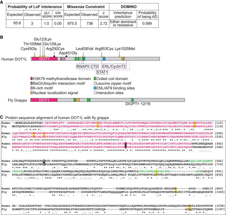
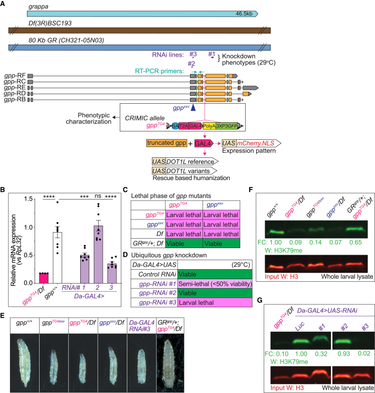
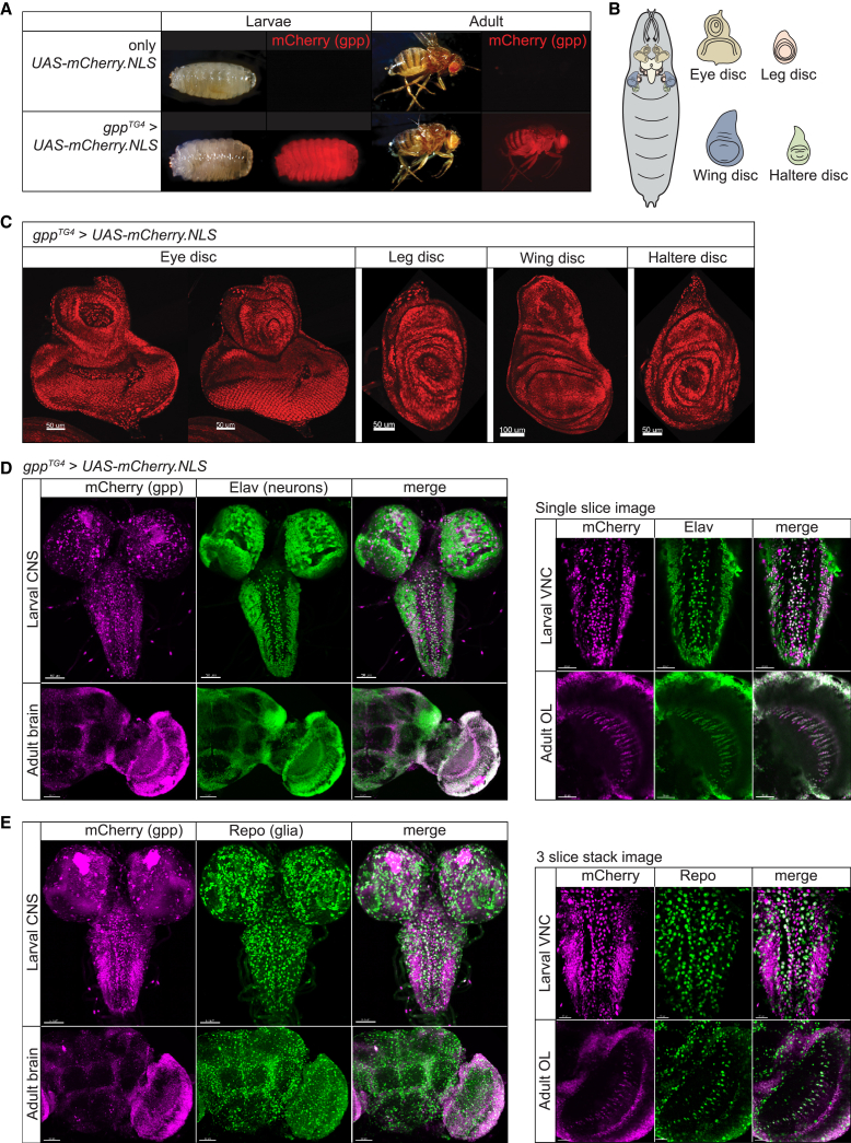
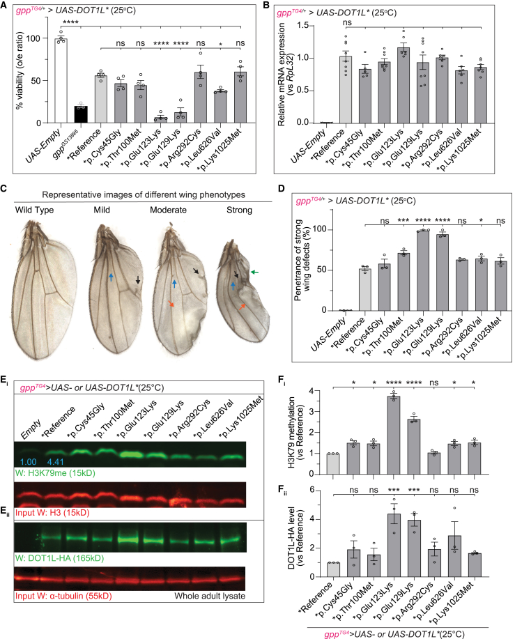
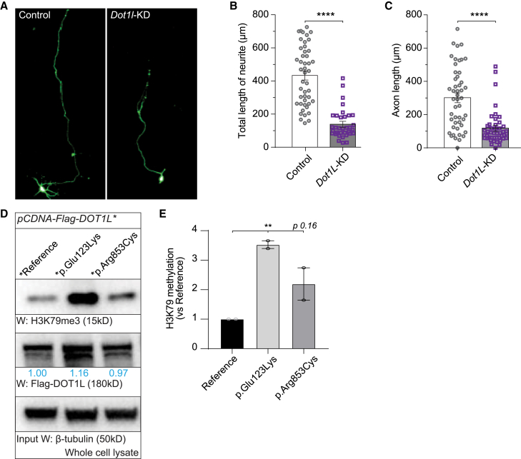
Misregulation of histone lysine methylation is associated with several human cancers and with human developmental disorders. DOT1L is an evolutionarily conserved gene encoding a lysine methyltransferase (KMT) that methylates histone 3 lysine-79 (H3K79) and was not previously associated with a Mendelian disease in OMIM. We have identified nine unrelated individuals with seven different de novo heterozygous missense variants in DOT1L through the Undiagnosed Disease Network (UDN), the SickKids Complex Care genomics project, and GeneMatcher. All probands had some degree of global developmental delay/intellectual disability, and most had one or more major congenital anomalies. To assess the pathogenicity of the DOT1L variants, functional studies were performed in Drosophila and human cells. The fruit fly DOT1L ortholog, grappa, is expressed in most cells including neurons in the central nervous system. The identified DOT1L variants behave as gain-of-function alleles in flies and lead to increased H3K79 methylation levels in flies and human cells. Our results show that human DOT1L and fly grappa are required for proper development and that de novo heterozygous variants in DOT1L are associated with a Mendelian disease.

Keywords: DOT1 Like histone lysine methyltransferase; DOT1L; Drosophila; H3K79 methylation; congenital anomalies; developmental delay; gain of function; gpp; grappa; histone lysine methyltransferase.


Conflict of interest statement

Declaration of interests The authors declare no competing interests.

Figures

Figure 1
Figure 1
DOT1L is loss of function constrained and conserved in flies (A) DOT1L is variant constrained based upon the control population database, gnomAD. Variants in DOT1L are predicted to be inherited “either recessive or dominant” and have a score of 0.599 for “probability of being autosomal dominant” by DOMINO. (B) Protein domain organization of human DOT1L and its fly (gpp) ortholog. Domains of DOT1L and the region they span specified with amino acid numbers are as follows: catalytic domain DOT1 (16–330),, Bat3/Ubiquitin interaction motif (361–380), K-rich motif (390–407) with a partially overlapping nuclear localization signal (NLS) (395–417), three coiled-coil domains (529–549, 564–595, 616–636), leucine zipper motif (576–594), CTD binding patch (618–627), three ENL/AF9 binding sites (628–653, 863–878, 877–900),,, two more NLSs (1,088–1,111 and 1,164–1,171), and STAT1 interaction region (580–1,138). Variants suspected to be diagnostic are indicated above the protein as yellow dots and the variants suspected to be non-diagnostic are indicated as black dots. DOT1L’s ortholog in fly, gpp, has a DOT1 domain, three coiled-coil domains, and two NLSs. Gpp has a homology score 12/16 (DIOPT, v.8.5). (C) Protein sequence alignment of human DOT1L (Uniprot: Q8TEK3) and fly gpp (UniProt: Q8INR6). The variants are highlighted with yellow or black boxes. Pink letters correspond to the DOT1 domain and green letters correspond to a coiled-coil domain. Symbols in the protein alignment: fully conserved (), strongly similar (:), weakly similar (.), absent (−).
Figure 2
Figure 2
Fly grappa is essential for development and loss of gpp leads to developmental delay and a reduction in H3K79 methylation (A) Gene structure of fly gpp, fly reagents, and assays used in this study. The gene region is shown in light blue. A deficiency and genomic rescue (GR) construct are shown in brown and dark blue, respectively. Three independent RNAi lines (#1, P{TRiP.JF01284}attP2; #2, P{TRiP.GL01325}attP2; and #3, P{TRiP.HMJ02129}attP40) tested in this study and their target regions are shown in purple. Different isoforms of gpp (RB, RC, RD, RE, and RF) are shown in the middle panel and exons are indicated in orange, introns as black lines, and untranslated regions (UTR) in gray. gpp mutants used in phenotypic characterization studies include gppxxv (X-ray mutagenesis) and gppTG4 (CRIMIC allele). (B) Relative gpp mRNA expression levels are lower than 20% in gppTG4/Df mutant larvae when compared to controls (gpp+/+) based on real-time qPCR using primers shown in (A). Real-time qPCR analysis of gpp mRNA levels in ubiquitous gpp knockdown larvae shows that Da-GAL4 > gpp-RNAi reduces gpp expression to different levels. Da-GAL4 > gpp-RNAi #1 decreases gpp mRNA levels by ∼50% and RNAi #3 by 70%. RNAi #2 does not have any effect on gpp levels. Normalized gpp levels from three independent experiments were plotted as mean ± SEM, and statistical significance was determined by one-way ANOVA for multiple groups (∗∗∗p < 0.001, ∗∗∗∗p < 0.0001). (C) Animals homozygous for gpp mutant alleles are larval lethal. None of the alleles or a large deficiency allele which lacks gpp, Df(3R)BSC193, can complement each other. One copy of a genomic rescue construct inserted in second chromosome (VK37), GRgpp/+, rescues lethality of trans-heterozygous allelic combinations. (D) Ubiquitous knockdown of gpp results in larval lethality. While Da-GAL4 > RNAi #3 flies are larval lethal, -RNAi #1 flies are semi-lethal, as shown by lower-than-expected genotypic ratios of survival into adulthood. RNAi #2 flies are completely viable. Da-GAL4 > UAS-gpp-RNAi flies were compared to Da-GAL4 > control-RNAi (control-RNAi = UAS-luciferase-RNAi). All the crosses were performed at 29°C. (E) Complete loss or ubiquitous knockdown (<50%) of gpp leads to severe developmental delay. Images of age-matched larvae at third instar larval stage is shown. One copy of a genomic rescue construct inserted in second chromosome (VK37), GRgpp/+, can rescue the developmental delay phenotype. (F) Gpp mutant animals have a drastic decrease in H3K79 methylation levels. Protein lysate from 10 larvae was prepared for each sample. H3K79 methylation levels were normalized with loading control, H3, and fold change (FC) for each sample were calculated by comparing normalized H3K79 methylation levels to wild-type larvae (gpp+/+). One copy of the genomic rescue construct, GRgpp/+, can increase the H3K79 methylation levels to ∼65%. (G) Ubiquitous knockdown (<50%) of gpp causes a severe decrease in H3K79 methylation levels. Da-GAL4 > gpp-RNAi #1 decreases H3K79 methylation levels to ∼30% and RNAi #3 to ∼2%. RNAi #2 does not have any effect on methylation levels. Protein lysate from 10 larvae was prepared for each sample. H3K79 methylation levels were normalized with loading control, H3, and FC for each sample were calculated by comparing normalized H3K79 methylation levels to control-RNAi (control-RNAi = UAS-luciferase-RNAi). All the crosses were performed at 29°C. A sample which is excluded from this study is cropped out from the gel image.
Figure 3
Figure 3
Fly grappa is broadly expressed in both larvae and adult flies The gppTG4 allele was used to drive expression of UAS-fluorescent reporter transgene (UAS-mCherry.NLS). (A) gpp is expressed broadly and highly throughout the body in both larvae and adult flies. Nuclear mCherry was present and whole larvae or adult was imaged. (B) Schematic of different tissues of larvae. (C) gpp is expressed in most tissues in larvae. Nuclear mCherry was present and tissue were dissected from L3 larvae including eye, leg, wing, and haltere discs. (D and E) gpp is broadly expressed in the nervous system. Nuclear mCherry was driven by gppTG4/+ and tissues were dissected from L3 larvae (CNS, includes central brain and VNC) or adults (brain). Shown is half of the adult brain. Tissue was counterstained with markers for neurons (Elav) (D) or glia (Repo) (E). Z stacked images of gpp expression pattern compared to neurons or glia are shown in the left panels in (D) and (E), respectively. Single slice images (right panels) were used to better visualize cellular co-localization of Nuclear mCherry signal with neurons (D) or glia (E). Note that gpp is expressed in a large subset of neurons and in a small subset of glia. VNC, ventral nerve cord; OL, optic lobe.
Figure 4
Figure 4
Expression human DOT1L disrupts wing morphology and different variants display different amounts of toxicity and H3K79 methylation (A) Heterozygous mutant flies, gppTG4/+, expressing reference or variant DOT1L cDNA have reduced viability compared to control flies (gppTG4/+> UAS-Empty) as shown by lower-than-expected genotypic ratios of survival into adulthood. The variants p.Glu123Lys and p.Glu129Lys show significant reduction in viability compared to flies expressing reference DOT1L. All the crosses were performed at 25°C. Percent viabilities (o/e ratios) from three independent experiments were plotted as mean ± SEM, and statistical significance was determined by one-way ANOVA for multiple groups (∗∗∗∗p < 0.0001). (B) Relative DOT1L mRNA expression levels in gppTG4/+ flies expressing reference or variant DOT1L. Normalized DOT1L levels from three independent experiments were plotted as mean ± SEM, and statistical significance was determined by one-way ANOVA for multiple groups. (C) Representative images of different wing phenotypes observed in in survivors of heterozygous mutant flies, gppTG4/+, expressing each DOT1L cDNA. Blue arrow, loss of cross-vein; black arrow, extra vein branching; orange arrow, blistered areas; green arrow, necrotic areas. (D) Penetrance of strong wing phenotypes in survivors of heterozygous mutant flies, gppTG4/+, expressing each DOT1L cDNA. Percent penetrance for strong phenotype from three independent experiments were plotted as mean ± SEM, and statistical significance was determined by one-way ANOVA for multiple groups (p < 0.05, ∗∗∗p < 0.001, ∗∗∗∗p < 0.0001). (E) All the DOT1L variants, except p.Arg292Cys, behave as gain-of-function (GoF) mutations. Survivors of heterozygous mutant flies, gppTG4/+, expressing reference or variant DOT1L cDNA show increased H3K79 methylation compared to control flies (gppTG4/+> UAS-Empty). Flies expressing variant DOT1L cDNAs, corresponding to p.Cys45Gly, p.Thr100Met p.Glu123Lys, p.Glu129Lys, p.Leu626Val, and p.Lys1025Met, show higher H3K79 methylation levels when compared to reference (Ei). Flies expressing variant DOT1L cDNAs p.Glu123Lys and p.Glu129Lys show higher DOT1L levels when compared to reference (Eii). Protein lysate from 10 adult flies were prepared for each sample. H3K79 methylation levels were normalized with loading control, H3, and fold change for each sample were calculated by comparing normalized H3K79 methylation levels to reference DOT1L-expressing flies. DOT1L levels were normalized with loading control, α-tubulin, and fold change for each sample were calculated by comparing normalized DOT1L levels to reference DOT1L-expressing flies. All the crosses were performed at 25°C. Blue numbers indicate the fold change of H3K79 methylation level in reference when compared to control (UAS-Empty). (Fi) Normalized H3K79 methylation band intensities for each group from three independent experiments were plotted as mean ± SEM, and statistical significance was determined by one-way ANOVA for multiple groups (p < 0.05, ∗∗∗∗p < 0.0001). (Fii) Normalized DOT1L band intensities for each group from three independent experiments were plotted as mean ± SEM, and statistical significance was determined by one-way ANOVA for multiple groups (∗∗∗p < 0.001).
Figure 5
Figure 5
Knockdown of Dot1l decreases axonal length and human DOT1L variants increase H3K79 methylation levels (A) Representative images of different primary neuronal cells. Left panel shows mouse primary neurons transfected with control plasmid and right panel shows mouse primary neurons transfected with Dot1l-shRNA plasmid. (B and C) Total neurite length (B) and total axonal length (C) was measured using ImageJ (n = 50 per condition). Results were plotted as mean ± SEM, and statistical significance was determined by unpaired t test (∗∗∗∗p < 0.0001). (D) DOT1L variants behave as gain-of-function (GoF) mutations. HEK293T cells were transfected with N-terminal flag tagged reference or variant (p.Glu123Lys and p.Arg853Cys) DOT1L cDNAs. Whole cell lysate was prepared for each sample. H3K79 methylation levels were normalized with loading control, β-tubulin, and fold change for each sample were calculated by comparing normalized H3K79 methylation levels to reference DOT1L-expressing cells. Blue numbers indicate the fold change of DOT1L levels in cells expressing variant compared to cells expressing reference DOT1L. (E) Normalized H3K79 methylation band intensities for each group from two independent experiments were plotted as mean ± SEM, and statistical significance was determined by one-way ANOVA for multiple groups (∗∗p < 0.01).

References

    1. Kouzarides T. Chromatin Modifications and Their Function. Cell. 2007;128:693–705. doi: 10.1016/j.cell.2007.02.005. - DOI - PubMed
    1. Greer E.L., Shi Y. Histone methylation: A dynamic mark in health, disease and inheritance. Nat. Rev. Genet. 2012;13:343–357. doi: 10.1038/nrg3173. - DOI - PMC - PubMed
    1. Husmann D., Gozani O. Histone lysine methyltransferases in biology and disease. Nat. Struct. Mol. Biol. 2019;26:880–889. doi: 10.1038/s41594-019-0298-7. - DOI - PMC - PubMed
    1. Allis C.D., Berger S.L., Cote J., Dent S., Jenuwien T., Kouzarides T., Pillus L., Reinberg D., Shi Y., Shiekhattar R., et al. New Nomenclature for Chromatin-Modifying Enzymes. Cell. 2007;131:633–636. doi: 10.1016/j.cell.2007.10.039. - DOI - PubMed
    1. Faundes V., Newman W.G., Bernardini L., Canham N., Clayton-Smith J., Dallapiccola B., Davies S.J., Demos M.K., Goldman A., Gill H., et al. Histone Lysine Methylases and Demethylases in the Landscape of Human Developmental Disorders. Am. J. Hum. Genet. 2018;102:175–187. doi: 10.1016/j.ajhg.2017.11.013. - DOI - PMC - PubMed

Publication types