. 2023 Nov 15:282:120406.
doi: 10.1016/j.neuroimage.2023.120406. Epub 2023 Oct 11.

Mapping mesoscale connectivity within the human hippocampus

Affiliations

Mapping mesoscale connectivity within the human hippocampus

Michel Modo et al. Neuroimage. .

Abstract

The connectivity of the hippocampus is essential to its functions. To gain a whole system view of intrahippocampal connectivity, ex vivo mesoscale (100 μm isotropic resolution) multi-shell diffusion MRI (11.7T) and tractography were performed on entire post-mortem human right hippocampi. Volumetric measurements indicated that the head region was largest followed by the body and tail regions. A unique anatomical organization in the head region reflected a complex organization of the granule cell layer (GCL) of the dentate gyrus. Tractography revealed the volumetric distribution of the perforant path, including both the tri-synaptic and temporoammonic pathways, as well as other well-established canonical connections, such as Schaffer collaterals. Visualization of the perforant path provided a means to verify the borders between the pro-subiculum and CA1, as well as between CA1/CA2. A specific angularity of different layers of fibers in the alveus was evident across the whole sample and allowed a separation of afferent and efferent connections based on their origin (i.e. entorhinal cortex) or destination (i.e. fimbria) using a cluster analysis of streamlines. Non-canonical translamellar connections running along the anterior-posterior axis were also discerned in the hilus. In line with "dentations" of the GCL, mossy fibers were bunching together in the sagittal plane revealing a unique lamellar organization and connections between these. In the head region, mossy fibers projected to the origin of the fimbria, which was distinct from the body and tail region. Mesoscale tractography provides an unprecedented systems view of intrahippocampal connections that underpin cognitive and emotional processing.

Keywords: Connectivity; Dentate Gyrus; Hippocampus; Mesoscale; Mossy Fibers; Perforant path; Tractography; Tri-synaptic circuit.


Conflict of interest statement

Declaration of Competing Interest The authors have no personal financial or institutional interest in the results described in this article.

Figures
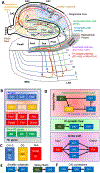
Fig. 1.
Fig. 1.. Hippocampal connectivity.
A. Schematic overview of hippocampal connectivity. Anatomical parcellation of a hippocampal slices of the body defines the classival view of its organization into subfields and cell layers, as well axonal connectivity. Extra-hippocampal connectivty through the di- and tri-synaptic path, as well as the temporoammonic path with the lateral (LEA) and medial entorhinal area (MEA) traverse through the subicular complex adjacent to the angular bundle. The tri-synaptic and temporoammonic path form the perforant path, the major fiber input to the hippocampus. The alevear path in contrast provides the main route of information output to the MEA, as well as the fimbria. Shorter distance intra-hippocampal connections are provided by Schaeffer collaterals and Mossy fibers. B. The hippocampus proper consists only of the 3 cornu ammonis (CA) subfields, whereas inclusion of the dentate gyrus (DG) and alveus (Alv) is commonly refered to as the hippocampus (HC). It also contains the hippocampal fissure (Fiss), but this does not contain any cells. The hippocampal formation further expands the hippocampus to include the Pro-Subiculum (ProS), Subiculum (Sub), Pre-Subiculum (PrS), Para-Subiculum (ParaS), Fimbria (Fim), and Uncus. Addition of the angular bundle (AB), entorhinal (EC) and peri-rhinal cortices (PRC) is often referred to as the para-hippocampal gyrus (PHG). C. The hippocampus is part of the allocortex contained 3 to 4 cell layers. Notably the CA1–3 subfield contain the stratum moleculare (SM), stratum radiatum (SR), stratum pyramidale (a.k.a. pyramidal cell layer, PCL) and arguably the stratum oriens (SO). The DG contains the SM, granuel cell layer (GCL) and the hilus. Contiguous with the CA subfields, the subiculum also is considered to have 3–4 cell layers consisting of the SM/SR, PCL and stratum polymorpha (SP). It provides the transition area to the 5 layered periallocortex (i.e. EC and PRC). D. Schematic representation of the temporoammonic “direct’ and tri-synpatic “indirect” path constituting the major hippocampual afferent known as the perforant path, as well as the di-synaptic loop, which also contributes afferent connections. The alveus serves as a gateway from efferent and afferent fibers. E. Schaeffer collaterals are part of the tri-synpatic path by connecting neurons from the CA3 subfield with those from the CA1 subfield (synapse 3), whereas Mossy fibers connect neurons in the GCL with the neurons in the CA3 subfield. F. The CA2 subfield provides a connectivity bridge between both CA3 and CA1 with direct connections to both subfields. However, anatomically the CA2 subfield is visually difficult to separate from both the CA1 and CA3 subfields.
Fig. 2.
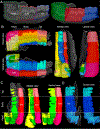
Fig. 2.. 3D renderings of hippocampal segmentations.
A. Hippocampal subregion outline and rendering illustrating the head, body and tail volumes. B. 3D renderings of subfields for the head, body and tail region illustrate how their volumes and shape changes, especially in the head region. C. Illustration of the PCL in CA1–3 and its relationship with the hilar region. It is noteworthy that the PCL present in a more consistent smoother fashion compared to the hilus, which exhibit the dentation pattern observed in the dentate gyrus. D. 3D renderings of the GCL in the head, body and tail region. Dentations along the body and tail region are evident in this rendering, as well as “horn” like structures in the head region.
Fig. 3.
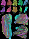
Fig. 3.. Hippocampal tractography.
A. Tractography of the entire hippocampal sample reveals a thorough mesh of connections. Schaffer collaterals in the PCL provide a unique view of this cell layer with the overlying cortex reveal connections running perpendicular to the PCL. The head region also stands out with the DG subfield showing a nexus of connections. Streamlines in the angular bundle run perpendicular to those in PCL, as well as those in the EC. B. Separation of streamlines by size achieves a unique view of short distance intra-hippocampal connections (<3 mm in length) to contrast these with long distance connections (>7 mm in length). C. A combined view of these different scales of connections provides novel insights into which parts of the hippocampus is dominated by intra-hippocampal connections versus those contain input and output connections from surrounding regions.
Fig. 4.
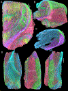
Fig. 4.. Cluster analysis of tractography.
A. An expectation maximizing (EM) algorithm created 10 clusters of streamlines that share particular charateristics to create a hypthosis-free view at hippocampal connectivity. The first 4 clusters contain 67.26% of all connections. The first two clusters mainly consist of short distance connections, which are commonly omitted in point-to-point analysis (i.e. virtual dissections) as they are contained within the same ROI. Smaller clusters (e.g clusters 9 and 10) encompass longer streamlines with separate anatomical substrates in the alveus. These could not easily be separated using distance of streamlines or creating separate ROIs based on anatomical location. B. Consolidated sagittal view of all color-coded clusters for the enitre hippocampal sample. C. Consolidated coronal view of a middle body slice reveals a separation of connections in the alveus to highlight those streamlines converging into the fimbria versus those merging with the angular bundle. D. By eliminating short distance connections in clusters 1–3, a skeleton view of hippocampal connectivity is visible that highlights how connectivity can be used to delineate subfields. Notably CA1 is characterized by the perforant path, whereas the ProS and Sub have a unique shape of PCL fibers compared to the PrS area.
Fig. 5.
Fig. 5.. Whole hippocampus connectivity.
A. Coronal and sagittal cut of a body slice reveals anterior-posterior connections in the hilar region, as well as the Mossy fibers connecting to these fibers. The perforant path and its composition of different tracts is also evident. Efferent and afferent fibers can be distinguished based on their anatomical location and traced adjacent to the angular bundle fibers. B. A major advantage of MRI is the volumetric nature of its data that affords a visualization of fibers in a 3D volume to gain a systems view, as well as how different fiber systems interact. C. A note in point is the alveus, which comprises different layers of axons. Clustering of connections in the whole hippocampus hence affords a unique visualization of all the streamlines in the alveus and how these overlap or separate according to their anatomical location. D. A surface view of the 3 fiber clusters found in the alveus and their juxtaposition with those of the angular bundle.
Fig. 6.
Fig. 6.. Connectivity of the granule cell layer.
A. 3D rendering of the GCL for the entire hippocampus and tracing of its connections. B. Separation of GCL-based tracts according to the hippocampal region. Anterior-posterior connections are mostly associated with the tail region and extent to the area of the fimbria. Bunches of fibers is very evident in the body and tail region, but not so in the head region.
Fig. 7.
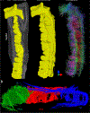
Fig. 7.. Connectivity of granule cell layer in the head sub-region.
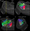
A. Coronal-sagittal view of the head region (yellow line = intersection point). The anatomical organization of head region is quite different from the body and tail region, especially for the granule cell layer and the hilar region. This is further reflected in the connectivity pattern with mossy fibers mostly projecting towards the emergence of the fimbria region, which is at the border of the head and body. B. The sagittal plane provides a clear indication of the mossy fibers from the GCL converging to the emergence of the fimbria from throughout the head region. Essentially, an extended space of connections are funnel towards a singular point to converge with the fimbria. C. In the coronal plane, the convergence of mossy fibers towards the fimbria is also evident, including dorsal GCL cells.
Fig. 8.
Fig. 8.. Connecivity of the pyramidal cell layer.
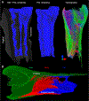
A. 3D rendering of the PCL reveals the smoother nature of this cell layer compared to the GCL, but also its hollow curvature. The head region stands out in its shape compared to the body and tail regions. Tractography further reveals that the head region has a unique organzation compared to the body and tail. Long anterior-posterior connections are also revealed and mostly associated with the input/output of fiber connections to the hippocampus through the PCL. B. Color-coded regional connections of the PCL in the sagittal plane further highlight how the PCL of the head region is a major contributor to the fimbria.
Fig. 9.
Fig. 9.. Canonical pathways – perforant path.
A. Cluster analysis of connections and selective visualization of these afforded a separation of individual well recognized pathways. Notably the tri-synpatic pathway could be visualized in a volumetric fashion rather than single cell tracings. It also affords a visualization of how the tri-synpatic and temporoammonic paths form the perforant path. The color-coding also aids in tracing these individual pathways as they merge with alvear fibers to define its anatomical position witihn this major fiber tract. A fiber tract supporting subicular fiber was also evident and terminates before the emergence of the perforant path from the alveus, further supporting this delineation as a border of the ProS and CA1. B.Visualization of the perforant path and its emergence from the alveus demonstrate how separation and combination of fiber tracts can aid in our understanding of how these systems interact. C. Direction-encoded coloring of fibers tracts in the temporoammonic path show how these funnel through CA1. D. Visualization of the first synpase in the tri-synpatic path as it emerges from the perforant path through CA1 into the DG.
Fig. 10.
Fig. 10.. Canonical pathways – alveus.
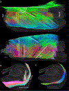
A. Different layers of fibers are overlapping in the alveus and these have a different angularity. However, fibers witihn each layer are well aligned with each other. Considering the destination of these fibers they can be distinguished as efferent and afferent. Afferent fibers from the perforant path appear to be more aligned with lamellar organization of the hippocampus. B. A coronal slice, representing the lamellar organization of the hippocampus, further illustrates the alignment of these perforant and subiciular path with a single slice. C. Color-coded direction of only the fibers in the alvear path also reveals a certain tortuosity of the tract between the subicular complex and angular bundle.
Fig. 11.
Fig. 11.. Non-canonical pathways –Anterior-posterior connectivity.
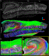
A. Tractography of the entire hippocampus revealed anterior-posterior connections running along the hilar region for the entire sample, including the head region. However, these connections were not very long fibers, but shorter (2–3 mm) long fibers that spanned across “dentations” and potentially defined functional lamellar units. Based on the appearance of these dentations, these functional units consist of different sizes and have a slightly varying shape of the GCL and hilar region. B. A closer view of these anterior-posterior connecting fibers also reveals within the “dentation” angle fibers that appear to integrate lamella in the sagittal plane. C. A further anterior-posterior fiber bundle appears in the hilar part of the crescent of the inner limb of the GCL. Both anterior-posterior connections are in close proximity with mossy fibers.
Fig. 12.
Fig. 12.. Lamellar bundling of mossy fibers.
A. Anterior-posterior running fibers are mostly located around the hilus, with mossy fibers running between these connections. B. Three main anterior-posterior connections can be seen within a lamella. C. A sagittal view reveals a bundling or bunch of mossy fibers with anterior-posterior connections providing a link between these units at either end.

References

    1. Adler DH, Pluta J, Kadivar S, Craige C, Gee JC, Avants BB, Yushkevich PA, 2014. Histology-derived volumetric annotation of the human hippocampal subfields in postmortem MRI. Neuroimage 84, 505–523. - PMC - PubMed
    1. Adler DH, Wisse LEM, Ittyerah R, Pluta JB, Ding SL, Xie L, Wang J, Kadivar S, Robinson JL, Schuck T, Trojanowski JQ, Grossman M, Detre JA, Elliott MA, Toledo JB, Liu W, Pickup S, Miller MI, Das SR, Wolk DA, Yushkevich PA, 2018. Characterizing the human hippocampus in aging and Alzheimer’s disease using a computational atlas derived from ex vivo MRI and histology. Proc. Natl. Acad. Sci. USA 115, 4252–4257. - PMC - PubMed
    1. Amaral DG, 1978. A Golgi study of cell types in the hilar region of the hippocampus in the rat. J. Comp. Neurol 182, 851–914. - PubMed
    1. Amaral DG, 1993. Emerging principles of intrinsic hippocampal organization. Curr. Opin. Neurobiol 3, 225–229. - PubMed
    1. Amaral DG, Witter MP, 1989. The three-dimensional organization of the hippocampal formation: a review of anatomical data. Neuroscience 31, 571–591. - PubMed

Publication types