

doi: 10.3174/ajnr.A8023.
Epub 2023 Oct 12.
A Novel Patient-Positioning Device for Dynamic CT Myelography
Affiliations
- PMID: 37827715
- PMCID: PMC10631527
- DOI: 10.3174/ajnr.A8023
Item in Clipboard
A Novel Patient-Positioning Device for Dynamic CT Myelography
AJNR Am J Neuroradiol.
2023 Nov.
Abstract
We describe a novel patient-positioning device for dynamic CT myelography. Dynamic CT myelography requires angling the patient's spine to distribute dense contrast along the dependent thecal sac. The proposed device is constructed of a low-density reinforced polymer frame and can be raised or lowered to various heights with a hand-operated mechanism, allowing precise adjustment of the spinal angle and control of the contrast bolus, increasing the safety, reproducibility, and sensitivity of dynamic CT myelography.
© 2023 by American Journal of Neuroradiology.
Figures

Spine angulation using a foam wedge versus using the elevation device in suspected ventral dural defects. A, A patient with a suspected ventral dural defect, positioned on top of a foam wedge for dCTM. CT image obtained after contrast injection shows pooling of contrast at the patient's lumbar lordosis (arrows) due to insufficient hip elevation. B, A separate patient with a suspected ventral dural defect on top of the novel positioning device. CT image obtained after contrast injection demonstrates egress of contrast from the puncture site to the craniocervical junction. C, Smaller FOV image demonstrates extravasation of contrast from the subarachnoid space into the ventral epidural space at C7–T1 (arrow), consistent with a ventral dural defect.
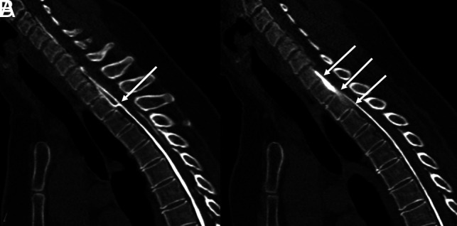
A ventral dural defect at T2–T3. A, Image obtained immediately after contrast injection demonstrates clear extravasation of contrast (arrow) from the subarachnoid space into the ventral epidural space, precisely localizing this patient's dural defect. B, A second image obtained 38 seconds after the first demonstrates rapid diffusion of contrast throughout the ventral epidural fluid collection (arrows), obscuring the precise site of the dural defect.

Device schematic.

Localization of a CSF venous fistula using the positioning device. Axial CT image with a wide FOV demonstrates a patient in the right lateral decubitus position on top of the positioning device during dCTM with a CSF venous fistula (solid arrows) arising from a right T8–T9 meningeal diverticulum. The device frame (dashed arrows) generates no streak artifacts.

Example of the device in use in decubitus (A, Flat. B, Elevated) and prone (C, Flat, D, Elevated) patient positions.
References
MeSH terms
LinkOut - more resources
Full Text Sources