Immuno-PET and Targeted α-Therapy Using Anti-Glypican-1 Antibody Labeled with 89Zr or 211At: A Theranostic Approach for Pancreatic Ductal Adenocarcinoma
- PMID: 37827841
- PMCID: PMC10690121
- DOI: 10.2967/jnumed.123.266313
Immuno-PET and Targeted α-Therapy Using Anti-Glypican-1 Antibody Labeled with 89Zr or 211At: A Theranostic Approach for Pancreatic Ductal Adenocarcinoma
Abstract
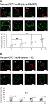
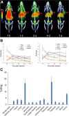
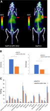
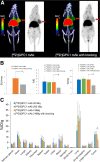
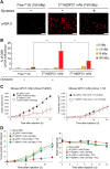
Glypican-1 (GPC1) is overexpressed in several solid cancers and is associated with tumor progression, whereas its expression is low in normal tissues. This study aimed to evaluate the potential of an anti-GPC1 monoclonal antibody (GPC1 mAb) labeled with 89Zr or 211At as a theranostic target in pancreatic ductal adenocarcinoma. Methods: GPC1 mAb clone 01a033 was labeled with 89Zr or 211At with a deferoxamine or decaborane linker, respectively. The internalization ability of GPC1 mAb was evaluated by fluorescence conjugation using a confocal microscope. PANC-1 xenograft mice (n = 6) were intravenously administered [89Zr]GPC1 mAb (0.91 ± 0.10 MBq), and PET/CT scanning was performed for 7 d. Uptake specificity was confirmed through a comparative study using GPC1-positive (BxPC-3) and GPC1-negative (BxPC-3 GPC1-knockout) xenografts (each n = 3) and a blocking study. DNA double-strand breaks were evaluated using the γH2AX antibody. The antitumor effect was evaluated by administering [211At]GPC1 mAb (∼100 kBq) to PANC-1 xenograft mice (n = 10). Results: GPC1 mAb clone 01a033 showed increased internalization ratios over time. One day after administration, a high accumulation of [89Zr]GPC1 mAb was observed in the PANC-1 xenograft (SUVmax, 3.85 ± 0.10), which gradually decreased until day 7 (SUVmax, 2.16 ± 0.30). The uptake in the BxPC-3 xenograft was significantly higher than in the BxPC-3 GPC1-knockout xenograft (SUVmax, 4.66 ± 0.40 and 2.36 ± 0.36, respectively; P = 0.05). The uptake was significantly inhibited in the blocking group compared with the nonblocking group (percentage injected dose per gram, 7.3 ± 1.3 and 12.4 ± 3.0, respectively; P = 0.05). DNA double-strand breaks were observed by adding 150 kBq of [211At]GPC1 and were significantly suppressed by the internalization inhibitor (dynasore), suggesting a substantial contribution of the internalization ability to the antitumor effect. Tumor growth suppression was observed in PANC-1 mice after the administration of [211At]GPC1 mAb. Internalization inhibitors (prochlorperazine) significantly inhibited the therapeutic effect of [211At]GPC1 mAb, suggesting an essential role in targeted α-therapy. Conclusion: [89Zr]GPC1 mAb PET showed high tumoral uptake in the early phase after administration, and targeted α-therapy using [211At]GPC1 mAb showed tumor growth suppression. GPC1 is a promising target for future applications for the precise diagnosis of pancreatic ductal adenocarcinoma and GPC1-targeted theranostics.
Keywords: astatine; glypican-1; immuno-PET; pancreatic ductal adenocarcinoma; targeted α-therapy; theranostics; zirconium.
© 2023 by the Society of Nuclear Medicine and Molecular Imaging.
Figures


References
-
- Miller KD, Siegel RL, Lin CC, et al. . Cancer treatment and survivorship statistics. CA Cancer J Clin. 2016;66:271–289. - PubMed
Publication types
MeSH terms
Substances
LinkOut - more resources
Full Text Sources
Medical
