Review
. 2023 Oct 13;8(1):396.
doi: 10.1038/s41392-023-01628-9.

Astrocytes in human central nervous system diseases: a frontier for new therapies

Affiliations
Review

Astrocytes in human central nervous system diseases: a frontier for new therapies

Alexei Verkhratsky et al. Signal Transduct Target Ther. .

Abstract

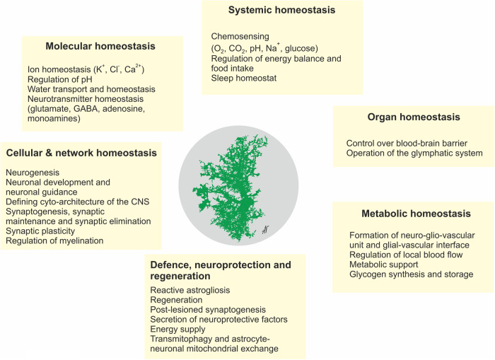
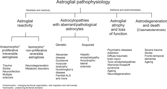
Astroglia are a broad class of neural parenchymal cells primarily dedicated to homoeostasis and defence of the central nervous system (CNS). Astroglia contribute to the pathophysiology of all neurological and neuropsychiatric disorders in ways that can be either beneficial or detrimental to disorder outcome. Pathophysiological changes in astroglia can be primary or secondary and can result in gain or loss of functions. Astroglia respond to external, non-cell autonomous signals associated with any form of CNS pathology by undergoing complex and variable changes in their structure, molecular expression, and function. In addition, internally driven, cell autonomous changes of astroglial innate properties can lead to CNS pathologies. Astroglial pathophysiology is complex, with different pathophysiological cell states and cell phenotypes that are context-specific and vary with disorder, disorder-stage, comorbidities, age, and sex. Here, we classify astroglial pathophysiology into (i) reactive astrogliosis, (ii) astroglial atrophy with loss of function, (iii) astroglial degeneration and death, and (iv) astrocytopathies characterised by aberrant forms that drive disease. We review astroglial pathophysiology across the spectrum of human CNS diseases and disorders, including neurotrauma, stroke, neuroinfection, autoimmune attack and epilepsy, as well as neurodevelopmental, neurodegenerative, metabolic and neuropsychiatric disorders. Characterising cellular and molecular mechanisms of astroglial pathophysiology represents a new frontier to identify novel therapeutic strategies.


Conflict of interest statement

Authors declare no competing interest. Alexey Semyanov, Alexei Verkhratsky and Peter Illes are the editors of STTT.

Figures

Fig. 1
Fig. 1
The nervous system with sensory input and motor output and multicellular active milieu of the nervous tissue. Reproduced from ref.
Fig. 2
Fig. 2
Diversity of astroglia
Fig. 3
Fig. 3
Functions of astroglia. The image of astrocyte is drawn based on 3D EM reconstruction kindly provided by Prof. Min Zhou, Ohio State University
Fig. 4
Fig. 4
Classification of astrogliopathology
Fig. 5
Fig. 5
Classification of reactive astrogliosis
Fig. 6
Fig. 6
Clasmatodendrosis of astrocytes. a Clasmatodendrosis as seen and drawn by Ramon y Cajal. A. Cell with preserved processes. B. Astrocyte with fragmentations. C, D, E. Astrocyte with disrupted cytoplasmic expansions, but with preservation of perikaryon. a. capillary. b. disaggregated end feet. b Clasmatodendrosis of astrocytes in the aged brain of mouse (stratum radiatum of dorsal hippocampus). Upper panel Astrocytes with distinctive enlarged soma and vacuolisation of processes distinctive to clasmatodendrosis for a representative cluster of astrocytes. Lower panel: Imaris surface render of a confocal z-stack of GFAP (blue), S100β (green), and Vimentin (Red) demonstrates an astrocyte with clasmatodendrosis (left) showing the co-localisation of S100β+Vimentin+ beads along GFAP+ processes, and a reactive astrocyte with non-degenerative morphology adjacent to it (right). Reproduced from ref.
Fig. 7
Fig. 7
Potential detrimental effects of diseased, atrophied, reactive or aged astrocytes that can occur in specific contexts. See text for further explanation
Fig. 8
Fig. 8
Stages of neuroinflammation and scar formation following traumatic brain injury. See text for explanation. Modified from ref.
Fig. 9
Fig. 9
Reactive astrogliosis and protective astrocyte border formation in experimental stroke. Images show an ischaemic infarct in mouse striatum at 14 days after injection of the vasoconstrictive agent, L-NIO (N-(1-Iminoethyl)-L-ornithine) in a manner similar to that described previously. Immunohistochemistry shows GFAP-expressing astrocytes stained red and NeuN-expressing neurones stained light blue. The left image shows the border of newly-proliferated reactive astrocytes that surround the fibrotic tissue (unstained) of the infarct core and isolate it from adjacent neural tissue. The right image shows a normal density of healthy neurones immediately adjacent to the protective astrocyte border. Images are courtesy of Dr. Shinong Wang and Dr. Yan Ao
Fig. 10
Fig. 10
Pathophysiology of the AQP4 form of the neuromyelitis optica. See text for explanation. Modified from ref.
Fig. 11
Fig. 11
Exposure to Chronic Unpredictable Stress (CUMS) induces morphological atrophy in prefrontal cortex astrocytes in mice. a Representative 3D reconstruction of astrocyte in control and CUMS groups. b Sholl analysis of astrocytic morphology for control and CUMS groups shows the number of intersections of astrocytic branches with concentric spheres centred in the middle of cell soma. c Maximal number of intersections for astrocytes in control and CUMS groups. d Average length of astrocytic processes in control and CUMS groups. bd n = 15 for each group. e Representative examples of astrocytic territorial domains obtained as a projection of astrocytes along the z-axis projection for control and CUMS animals. f Average astrocytic domain area (E) and average length of astrocytic processes for control and CUMS group. All data are presented as mean ± s.e.m. *p < 0.05, **p < 0.01, ***p < 0.001. The number of experiments is indicated on each column. Reproduced from ref.
Fig. 12
Fig. 12
Astrocytic atrophy in ageing. Upper panel shows 3D reconstructions of Alexa 594 filled cortical astrocytes of mice of different ages. Reproduced from ref.  Lower panel shows 3D of Alexa 594 filled astrocytes from the cortex of human patients (tissue obtained during neurosurgery) of different ages. Reproduced from ref.
Fig. 13
Fig. 13
Decline in astrocytic functions in ageing
Fig. 14
Fig. 14
Glial reactivity, decline and paralysis define the pathophysiology of AD. At the prodromal and early stages of the disease astrocytes display atrophic morphology possibly indicating limited homoeostatic support, which may lead to the early synaptic malfunction. After emergence of the plaques reactive astrocytes and microglia surround β-amyloid depositions to protect the nervous tissue. Progressive decline in glial neuroprotection and homoeostatic support culminates in glial paralysis which permits neuronal death and brain atrophy manifested in dementia
Fig. 15
Fig. 15
Histopathology of astrocytes in astrotauopathies. See text for explanation. Modified, and reproduced from the images kindly provided by Professor Gabor Kovacs, Toronto University

References

    1. Semyanov A, Verkhratsky A. Astrocytic processes: from tripartite synapses to the active milieu. Trends Neurosci. 2021;44:781–792. - PubMed
    1. Verkhratsky A, Arranz AM, Ciuba K, Pekowska A. Evolution of neuroglia. Ann. N.Y. Acad. Sci. 2022;1518:120–130. - PubMed
    1. Verkhratsky A, Nedergaard M. The homeostatic astroglia emerges from evolutionary specialization of neural cells. Philos. Trans. R. Soc. Lond. B Biol. Sci. 2016;371:20150428. - PMC - PubMed
    1. Verkhratsky A, Parpura V, Vardjan N, Zorec R. Physiology of astroglia. Adv. Exp. Med. Biol. 2019;1175:45–91. - PMC - PubMed
    1. Kettenmann, H. & Ransom, B. R. (Oxford University Press, Oxford, 2013).

Publication types

MeSH terms