. 2023 Nov;20(11):1379-1392.
doi: 10.1038/s41423-023-01088-9. Epub 2023 Oct 12.

A de novo missense mutation in MPP2 confers an increased risk of Vogt-Koyanagi-Harada disease as shown by trio-based whole-exome sequencing

Affiliations

A de novo missense mutation in MPP2 confers an increased risk of Vogt-Koyanagi-Harada disease as shown by trio-based whole-exome sequencing

Xianyang Liu et al. Cell Mol Immunol. 2023 Nov.

Abstract

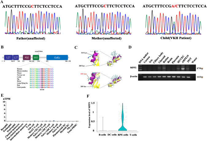
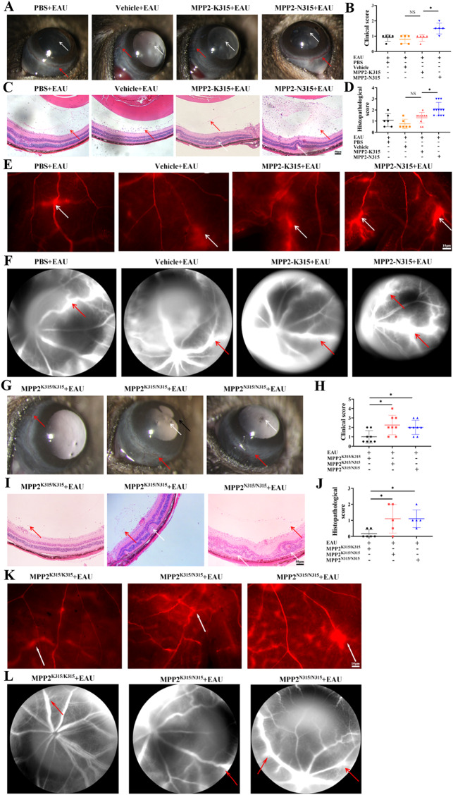
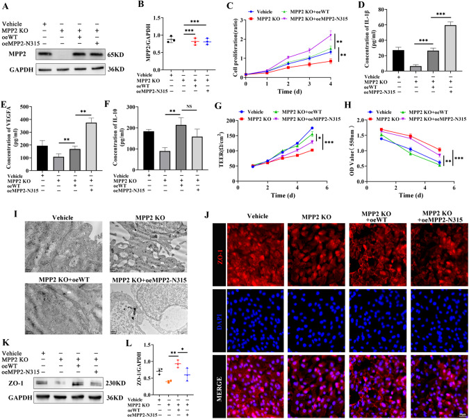
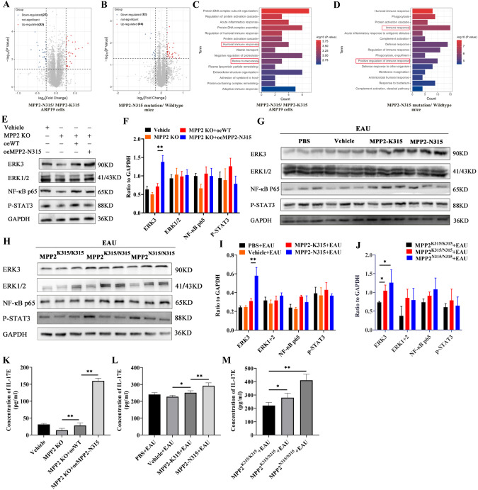
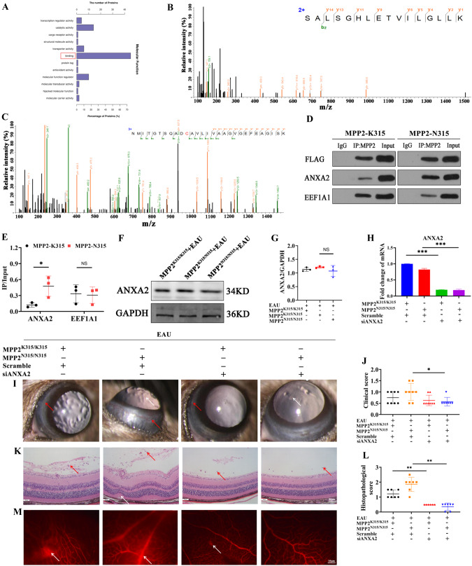
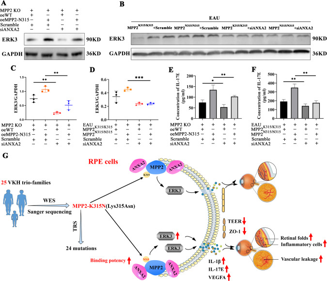
Vogt-Koyanagi-Harada (VKH) disease is a leading cause of blindness in young and middle-aged people. However, the etiology of VKH disease remains unclear. Here, we performed the first trio-based whole-exome sequencing study, which enrolled 25 VKH patients and 50 controls, followed by a study of 2081 VKH patients from a Han Chinese population to uncover detrimental mutations. A total of 15 de novo mutations in VKH patients were identified, with one of the most important being the membrane palmitoylated protein 2 (MPP2) p.K315N (MPP2-N315) mutation. The MPP2-N315 mutation was highly deleterious according to bioinformatic predictions. Additionally, this mutation appears rare, being absent from the 1000 Genome Project and Genome Aggregation Database, and it is highly conserved in 10 species, including humans and mice. Subsequent studies showed that pathological phenotypes and retinal vascular leakage were aggravated in MPP2-N315 mutation knock-in or MPP2-N315 adeno-associated virus-treated mice with experimental autoimmune uveitis (EAU). In vitro, we used clustered regularly interspaced short palindromic repeats (CRISPR‒Cas9) gene editing technology to delete intrinsic MPP2 before overexpressing wild-type MPP2 or MPP2-N315. Levels of cytokines, such as IL-1β, IL-17E, and vascular endothelial growth factor A, were increased, and barrier function was destroyed in the MPP2-N315 mutant ARPE19 cells. Mechanistically, the MPP2-N315 mutation had a stronger ability to directly bind to ANXA2 than MPP2-K315, as shown by LC‒MS/MS and Co-IP, and resulted in activation of the ERK3/IL-17E pathway. Overall, our results demonstrated that the MPP2-K315N mutation may increase susceptibility to VKH disease.

Keywords: Annexin A2; De novo mutation; ERK3/IL-17E pathway; Membrane palmitoylated protein 2; Vogt–Koyanagi–Harada disease; Whole exome sequencing.


Conflict of interest statement

All authors declare no competing interests.

Figures

Fig. 1
Fig. 1
Identification of de novo mutations for VKH disease. A Identification of the de novo MPP2 p.K315N mutation in one VKH trio family. B The predicted conservation for the MPP2-K315N mutation in humans, Rhesus monkeys, mice, etc. C The structure prediction of the MPP2-K315 and MPP2-N315 proteins. D The expression profile of the MPP2 gene in several human tissues and cell lines. E The nTPM of MPP2 in immune cells. F The expression levels of MPP2 in B cells, DC cells, RPE cells and T cells
Fig. 2
Fig. 2
Aggravation of inflammatory response and destruction of the BRB in MPP2-N315 mutant mice with EAU. A, B Anterior segment inflammation and clinical scores of mice with EAU treated with PBS, vehicle, MPP2 wild-type or MPP2-N315 mutant AAV. White arrow, inflammatory cells. Red arrow, conjunctival and ciliary hyperemia (n = 5/group; mean ± SD; NS p > 0.05, *p < 0.05; Mann–Whitney U test). C, D Hematoxylin-eosin staining and pathological scores of immunized mice with PBS, vehicle, MPP2 wild-type or MPP2-N315 mutant AAV. Red arrow, inflammatory cells. White arrow, retina fold (scale bar, 50 μm) (n = 6–12/group; mean ± SD; NS p > 0.05, *p < 0.05, **p < 0.01; Mann–Whitney U test). E Representative retinal flat-mount images of Evans blue on Day 14 after immunization in the four groups mentioned above. White arrow, vessel leakage (scale bar, 50 μm). F Representative images of fluorescein fundus angiography in the above four groups. Red arrow, vessel leakage. G, H Anterior segment inflammation and clinical scores of EAU in knock-in mice with MPP2-K315 or heterozygous or homozygous MPP2-N315 mutation. White arrow, inflammatory cells. Red arrow, conjunctival and ciliary hyperemia. Black arrow, posterior synechiae (n = 8/group; mean ± SD; *p < 0.05; Mann–Whitney U test). I, J Hematoxylin-eosin staining and histopathological scores in MPP2-K315 or heterozygous or homozygous MPP2-N315 knock-in mice with EAU. Red arrow, inflammatory cells. White arrow, retina fold (scale bar, 50 μm) (n = 5–6/group; mean ± SD; *p < 0.05; Mann–Whitney U test). K Images of Evans blue assays in the three groups mentioned above. White arrow, vessel leakage (scale bar, 10 μm). L Pictures of fluorescein fundus angiography in the three groups. Red arrow, vessel leakage
Fig. 3
Fig. 3
The effects of MPP2 mutation in ARPE19 cells with MPP2 deletion. A, B The KO efficiency of MPP2 and overexpression level of MPP2 wild-type or the MPP2 mutant (n = 3/group; mean ± SD; ***p < 0.001; one-way ANOVA). C The proliferation of ARPE19 cells with vehicle, MPP2 KO or overexpression of MPP2 mutant lentiviruses after MPP2 deletion (n = 5/group; mean ± SD; **p < 0.01; one-way ANOVA). DF The secretion levels of inflammatory factors including IL-1β, VEGFA and IL-10 by ELISA kits (n = 6/group; mean ± SD; NS > 0.05, **p < 0.01, ***p < 0.001; one-way ANOVA). G The electrical resistance of ARPE19 cells with vehicle, MPP2 KO or overexpression of MPP2 mutant lentiviruses after MPP2 deletion (n = 3/group; mean ± SD; *p < 0.05, ***p < 0.001; one-way ANOVA). H The phenol red leakage of ARPE19 cells with vehicle, MPP2 KO or overexpression of MPP2 mutant lentiviruses after MPP2 KO (n = 5/group; mean ± SD; **p < 0.01, ***p < 0.001; one-way ANOVA). I Images of transmission electron microscope (TEM) in the four groups mentioned above (scale bar, 1 μm). J The immunofluorescence results of ZO-1 (scale bar, 50 μm). K, L The protein expression and quantification of ZO-1 (n = 3/group; mean ± SD; *p < 0.05, **p < 0.01; one-way ANOVA)
Fig. 4
Fig. 4
Increased expression of inflammatory-related proteins after MPP2-N315 mutation. A The volcanic map of DEGs in MPP2-K315 or MPP2-N315 mutant ARPE19 cells. B The volcanic map of DEGs in MPP2 wild-type or mutant mice with EAU. C, D GO enrichment analysis of proteomics in vitro and in vivo. E, F Protein levels and quantitative charts of ERK3, ERK1/2, NF-κB p65, and p-STAT3 in vitro (n = 3/group; mean ± SD; **p < 0.01; one-way ANOVA). G, I Protein expression and quantification of ERK3, ERK1/2, NF-κB p65 and p-STAT3 in PBS, vehicle, MPP2-K315 or MPP2-N315 AAV-administered mice with EAU modeling (n = 3/group; mean ± SD; ** p < 0.01; one-way ANOVA). H, J Protein levels and quantitative graphs of ERK3, ERK1/2, NF-κB p65 and p-STAT3 in MPP2-K315 or homozygous or heterozygous MPP2-N315 knock-in mice with immunization (n = 3/group; mean ± SD; *p < 0.05; one-way ANOVA). K The extracellular protein level of IL-17E (n = 6/group; mean ± SD; **p < 0.01; one-way ANOVA). L The secretion level of IL-17E in mice with EAU treated with PBS, vehicle, MPP2-K315 or MPP2-N315 AAV (n = 6/group; mean ± SD; *p < 0.05, **p < 0.01; one-way ANOVA). M The secretion of IL-17E in MPP2-K315 or homozygous or heterozygous MPP2-N315 knock-in mice with immunization (n = 6/group; mean ± SD; *p < 0.05, **p < 0.01; one-way ANOVA)
Fig. 5
Fig. 5
Alleviated EAU inflammation after ANXA2 silencing. A Molecular function of MPP2 mutant in proteomics. B The top Ion score of the secondary spectrum of ANXA2. C The top Ion score of the secondary spectrum of EEF1A1. D Co-IP results of MPP2 to ANXA2 or EEF1A1. E The grayscale statistics of upper Co-IP results (n = 3/group; mean ± SD; NS p > 0.05, *p < 0.05; unpaired Student’s t test). F The protein level of ANXA2 in MPP2-K315 or homozygous or heterozygous MPP2-N315 knock-in mice with EAU. G Quantification of ANXA2 in the three groups mentioned above (n = 3/group; mean ± SD; NS > 0.05; one-way ANOVA). H Knockdown efficiency of ANXA2 in MPP2-K315 or MPP2-N315 knock-in mice with siANXA2 (n = 3/group; mean ± SD; ***p < 0.001; one-way ANOVA). I, J Clinical scores of MPP2-K315 or MPP2-N315 knock-in mice with or without ANXA2 silencing under immunization. White arrow, inflammatory cells. Red arrow, conjunctival and ciliary hyperemia (n = 8/group; mean ± SD; *p < 0.05; Mann–Whitney U test). K, L Histopathological scores in the four groups mentioned above. Red arrow, inflammatory cells. White arrow, retina fold (scale bar, 50 μm.) (n = 6–7/group; mean ± SD; **p < 0.01; Mann–Whitney U test). M Representative images of Evans blue in the above groups. White arrow, vessel leakage (scale bar, 10 μm)
Fig. 6
Fig. 6
Exacerbation of EAU inflammatory phenotypes via the MPP2/ANXA2/ERK3/IL-17E pathway. A, C The protein level and quantification of ERK3 in oeWT or oeMPP2-N315 ARPE19 cells with or without siANXA2 based on MPP2 KO (n = 3/group; mean ± SD; **p < 0.01; one-way ANOVA). B, D The expression and quantitative chart of ERK3 in MPP2-K315 or MPP2-N315 knock-in mice with scramble or siANXA2. (n = 3/group; mean ± SD; ***p < 0.001; one-way ANOVA). E The extracellular protein level of IL-17E (n = 6/group; mean ± SD; *p < 0.05, **p < 0.01; one-way ANOVA). F The secretion level of IL-17E in vivo (n = 6/group; mean ± SD; **p < 0.01; one-way ANOVA). G Schematic diagram of the MPP2-N315 mutation exacerbating EAU inflammation through the ANXA2/ERK3/IL-17E pathway

Similar articles

Cited by

References

    1. Rao NA. Uveitis in developing countries. Indian J Ophthalmol. 2013;61:253–4. doi: 10.4103/0301-4738.114090. - DOI - PMC - PubMed
    1. Tsirouki T, Dastiridou A, Symeonidis C, Tounakaki O, Brazitikou I, Kalogeropoulos C, et al. A focus on the epidemiology of uveitis. Ocul Immunol Inflamm. 2018;26:2–16. doi: 10.1080/09273948.2016.1196713. - DOI - PubMed
    1. Du L, Kijlstra A, Yang P. Vogt-Koyanagi-Harada disease: novel insights into pathophysiology, diagnosis and treatment. Prog Retin Eye Res. 2016;52:84–111. doi: 10.1016/j.preteyeres.2016.02.002. - DOI - PubMed
    1. Hou S, Li N, Liao X, Kijlstra A, Yang P. Uveitis genetics. Exp Eye Res. 2020;190:107853. doi: 10.1016/j.exer.2019.107853. - DOI - PubMed
    1. Yang P, Zhang Z, Zhou H, Li B, Huang X, Gao Y, et al. Clinical patterns and characteristics of uveitis in a tertiary center for uveitis in China. Curr Eye Res. 2005;30:943–8. doi: 10.1080/02713680500263606. - DOI - PubMed

Publication types