. 2023 Nov;30(11):2432-2445.
doi: 10.1038/s41418-023-01230-0. Epub 2023 Oct 12.

Ionizing radiation-induced mitophagy promotes ferroptosis by increasing intracellular free fatty acids

Affiliations

Ionizing radiation-induced mitophagy promotes ferroptosis by increasing intracellular free fatty acids

Pengfei Yang et al. Cell Death Differ. 2023 Nov.

Abstract

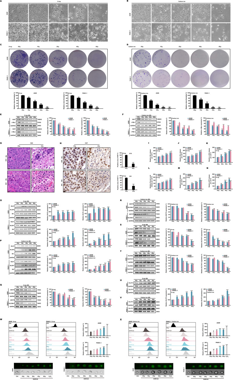
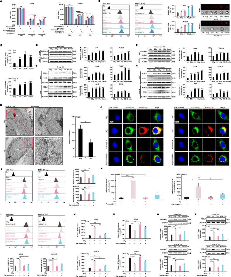
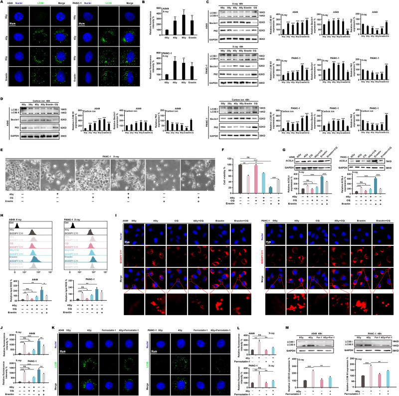
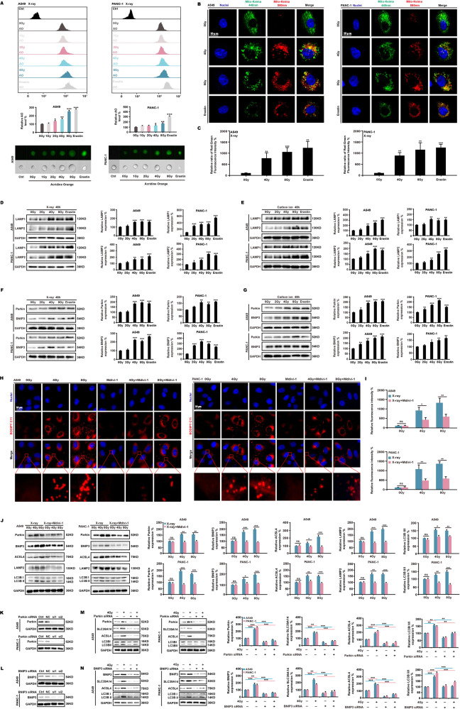
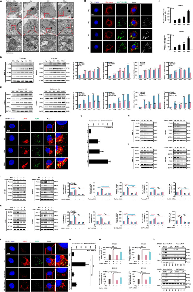
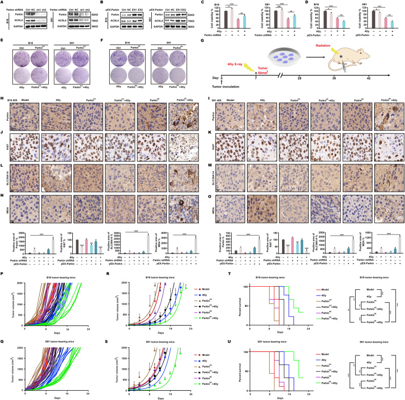
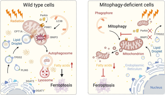
Ferroptosis is a type of cell death characterized by the accumulation of intracellular iron and an increase in hazardous lipid peroxides. Ferroptosis and autophagy are closely related. Ionizing radiation is a frequently used cancer therapy to kill malignancies. We found that ionizing radiation induces both ferroptosis and autophagy and that there is a form of mutualism between the two processes. Ionizing radiation also causes lipid droplets to form in proximity to damaged mitochondria, which, through the action of mitophagy, results in the degradation of the peridroplet mitochondria by lysosomes and the consequent release of free fatty acids and a significant increase in lipid peroxidation, thus promoting ferroptosis. Ionizing radiation has a stronger, fatal effect on cells with a high level of mitophagy, and this observation suggests a novel strategy for tumor treatment.


Conflict of interest statement

The authors declare no competing interests.

Figures

Fig. 1
Fig. 1. Ionizing radiation increases intracellular ROS.
A549 and PANC-1 cells were irradiated with X-ray and carbon ion beams. At 48 h, the morphology (A, B), and clone formation rate (C, D) were evaluated. Ki67 and c-Myc were identified in A549 and PANC-1 cells (E, F); H&E staining and ki67 expression were also performed in B16 and S91 melanomas (G, H). ELISA was used to assess glutamine (IK) and glycolytic (LN) metabolites. Western blotting was used to indicate glutaminase 1, glutamate dehydrogenase 1, glutamine synthetase (O, P), glutamine (Q, R) and glutamate (S, T) transport proteins and glucose transport proteins (U, V). Intracellular ROS production (W, X) was measured by flow cytometry.
Fig. 2
Fig. 2. Ionizing radiation induces ferroptosis.
A549 and PANC-1 cells were exposed to X-ray radiation and treated with various cell death inhibitor. At 48 h, it was found that ferrostatin-1 (2 μM) was effective in restoring cell survival (A). Using BODIPY C11 staining (1 μM), the amounts of lipid ROS generated by ionizing radiation were determined (B). Real-time PCR (C) and western blotting (DG) were used to assess the expression of major ferroptosis markers. The morphological changes induced in mitochondria by ionizing radiation were observed by transmission electron microscopy (H). Using flow cytometry and immunofluorescence, the levels of ROS (I), lipid peroxides (JM), and GPX4 (N) were determined. Eventually, the expression of critical ferroptosis proteins (O, P) was indicated.
Fig. 3
Fig. 3. Ferroptosis and autophagy are reciprocally influenced by ionizing radiation.
At 48 h of X-ray irradiation、erastin (5 μM) or CQ (2 μM) treatment, the expression of autophagy-related proteins in A549 and PANC-1 was assessed using immunofluorescence (A, B) and western blotting (C, D). The autophagy inhibitor CQ was used to treat the cells with X-ray radiation and erastin, and it was found that CQ restored cell survival (E, F). Through the measurement of ACSL4 (G), lipid ROS (H), and lipid peroxides (I, J), it was determined that CQ prevented ferroptosis. In addition, ferrostatin-1 (2 μM) combined with X-ray radiation inhibited the expression of the autophagy-essential protein LC3B (KM).
Fig. 4
Fig. 4. Mitophagy induced by ionizing radiation promotes ferroptosis.
A549 and PANC-1 cells were exposed to X-ray radiation and erastin (5 μM). At 48 h, AO staining (15 μg/mL) was used to identify intracellular acidic bodies (A). Using the Mito-Keima model, the shift of fluorescence was detected (B, C). In addition, the expression of lysosomal proteins (D, E) and mitophagy proteins (F, G) was evaluated. Immunofluorescence was used to investigate the formation of lipid peroxides (H, I) and the expression of ferroptosis- and mitophagy-related proteins (J) following X-ray radiation of cells with Mdivi-1 (2 μM). We then used siRNA to inhibit the expression of the essential genes of mitophagy (K, L), and X-ray radiation was used to assess the expression of the key proteins of ferroptosis (M, N).
Fig. 5
Fig. 5. Free fatty acids released by mitophagy boost lipid peroxidation.
At 48 h after irradiation and RSL3 (4 μM) treatment, transmission electron microscopy (A) and immunofluorescence (B) revealed the formation and aggregation of lipid droplets. The quantity of free fatty acids in PANC-1 and SW1990 cells was then determined using ELISA (C), and the expression of proteins involved in the production and localization of lipid droplets (D, E) was determined using western blotting. The colocalization of lipid droplets and lysosomes was subsequently examined (F, G). ParkinKD and BNIP3KD cells irradiated with X-ray radiation were used to assess the expression of lipid drop-related proteins (HK), and immunofluorescence was used to examine the colocalization of lysosomes and lipid drops (L, M). We used siRNA to inhibit the mitophagy genes in order to detect the quantity of free fatty acids (N). Finally, the co-immunoprecipitation approach was used to confirm the relationship between mitophagy and proteins involved in lipid droplet production and localization (O).
Fig. 6
Fig. 6. Ionizing radiation boosts killing in mitophagy-activated tumors through ferroptosis in mice.
Using shRNA and pEX-Parkin plasmids, stable B16 and S91ParkinKO cells (A) and ParkinOE cells (B) were created. At 48 h after irradiation, both cell survival (C, D) and clone formation (E, F) were identified. C57BL/6 mice were injected subcutaneously with B16 and S91 cells when tumor volume reached 50 mm3, then locally treated with a 4 Gy X-ray dose (G). After 48 h, tumors were harvested and assessed for signs of malignant proliferation and ferroptosis by immunohistochemistry (HO). On the remaining animals, tumor size was evaluated every two days following irradiation, and growth (PS) and survival (T, U) curves were plotted.
Fig. 7
Fig. 7. Schematic of the proposed mechanism.
Ionizing radiation induces substantial ROS in mitochondria, resulting in mitochondrial damage. Rapidly formed lipid droplets, under radiation stress, concentrate around damaged mitochondria to transfer fatty acids there. Mitophagy is simultaneously induced. Damaged mitochondria and lipid droplets form autolysosomes by lysosomal phagocytosis. Autolysosomes release free fatty acids into the cytoplasm, and after peroxidation by ROS, ferroptosis is strongly enhanced. This behavior does not occur in mitophagy-deficient cells; thus ionizing radiation-induced mitophagy promotes ferroptosis by increasing intracellular free fatty acid release.

Similar articles

Cited by

References

    1. Dixon SJ, Lemberg KM, Lamprecht MR, Skouta R, Zaitsev EM, Gleason CE, et al. Ferroptosis: an iron-dependent form of nonapoptotic cell death. Cell. 2012;149:1060–72. - PMC - PubMed
    1. Liang C, Zhang X, Yang M, Dong X. Recent Progress in Ferroptosis Inducers for Cancer Therapy. Adv Mater (Deerfield Beach, Fla) 2019;31:e1904197. - PubMed
    1. Ding H, Chen S, Pan X, Dai X, Pan G, Li Z, et al. Transferrin receptor 1 ablation in satellite cells impedes skeletal muscle regeneration through activation of ferroptosis. J cachexia, Sarcopenia Muscle. 2021;12:746–68. - PMC - PubMed
    1. Yu Y, Jiang L, Wang H, Shen Z, Cheng Q, Zhang P, et al. Hepatic transferrin plays a role in systemic iron homeostasis and liver ferroptosis. Blood. 2020;136:726–39. - PMC - PubMed
    1. Salnikow K. Role of iron in cancer. Semin Cancer Biol. 2021;76:189–94. - PubMed

Publication types

LinkOut - more resources