. 2023 Nov;42(47):3491-3502.
doi: 10.1038/s41388-023-02853-w. Epub 2023 Oct 12.

S1PR1 regulates ovarian cancer cell senescence through the PDK1-LATS1/2-YAP pathway

Affiliations

S1PR1 regulates ovarian cancer cell senescence through the PDK1-LATS1/2-YAP pathway

Yi-Ping Tao et al. Oncogene. 2023 Nov.

Abstract

Cell senescence deters the activation of various oncogenes. Induction of senescence is, therefore, a potentially effective strategy to interfere with vital processes in tumor cells. Sphingosine-1-phosphate receptor 1 (S1PR1) has been implicated in various cancer types, including ovarian cancer. The mechanism by which S1PR1 regulates ovarian cancer cell senescence is currently elusive. In this study, we demonstrate that S1PR1 was highly expressed in human ovarian cancer tissues and cell lines. S1PR1 deletion inhibited the proliferation and migration of ovarian cancer cells. S1PR1 deletion promoted ovarian cancer cell senescence and sensitized ovarian cancer cells to cisplatin chemotherapy. Exposure of ovarian cancer cells to sphingosine-1-phosphate (S1P) increased the expression of 3-phosphatidylinositol-dependent protein kinase 1 (PDK1), decreased the expression of large tumor suppressor 1/2 (LATS1/2), and induced phosphorylation of Yes-associated protein (p-YAP). Opposite results were obtained in S1PR1 knockout cells following pharmacological inhibition. After silencing LATS1/2 in S1PR1-deficient ovarian cancer cells, senescence was suppressed and S1PR1 expression was increased concomitantly with YAP expression. Transcriptional regulation of S1PR1 by YAP was confirmed by chromatin immunoprecipitation. Accordingly, the S1PR1-PDK1-LATS1/2-YAP pathway regulates ovarian cancer cell senescence and does so through a YAP-mediated feedback loop. S1PR1 constitutes a druggable target for the induction of senescence in ovarian cancer cells. Pharmacological intervention in the S1PR1-PDK1-LATS1/2-YAP signaling axis may augment the efficacy of standard chemotherapy.


Conflict of interest statement

The authors declare no competing interests.

Figures

Fig. 1
Fig. 1. Expression of S1PR1 in ovarian cancer tissues and paracancerous tissues, various organs, and ovarian cancer cell lines.
A Immunohistochemical analysis of S1PR1 in human ovarian cancer tissues (n = 6) and adjacent tissues (n = 2). Scale bar, 200µm (10 ×); Scale bar, 50µm (40 ×). B Quantitative analysis of immunohistochemical results. Student’s t-test; ***p < 0.001. C Western blot analysis of S1PR1 in human ovarian cancer tissues (CT) (n = 4) and paracancerous tissues (PT) (n = 2; left) and relative quantitative analysis (right). One-way ANOVA; ***p < 0.001. GAPDH was used as control. D Immunofluorescence analysis of S1PR1 in ovarian cancer cells. DAPI was used to counterstain nuclei (blue). Scale bar, 20µm. E Western blot analysis of S1PR1 in GC (granular cells derived from normal ovaries), ovarian cancer cell lines (ES-2, A2780, OVCAR-3, HO8910, SKOV3), and IOSE (ovarian cancer epithelial cell line; left) and relative quantitative analysis (right). GAPDH was used as control. One-way ANOVA; ns, p > 0.05; *p < 0.05; ***p < 0.001. F Western blot analysis of S1PR1 in organs (liver, lung, heart, kidney, tummy, skin, ovary, and testicle). GAPDH was used as control.
Fig. 2
Fig. 2. S1PR1 knockout affects the proliferation, migration, and the cell cycle of ovarian cancer cells.
A Western blot analysis of S1PR1 expression in wild-type and S1PR1 knockout ovarian cancer cells (A2780 and ES-2). One-way ANOVA; ***p < 0.001. β-actin was used as control. B Cell counting assay was used to detect the proliferation ability of wild-type ovarian cells and S1PR1 knockout cells (A2780 and ES-2). Counting was performed on days 1, 2, and 3 after inoculation and entailed 3 technical replicates per sample. One-way ANOVA; ***p < 0.001. C Soft agar cloning assay was used to compare the proliferation ability of wild-type ovarian cancer cells and S1PR1 knockout cells. Values represent mean ± SD of three independent experiments. One-way ANOVA; *p < 0.05, ***p < 0.001. D Transwell assay analysis of the migration ability of S1PR1 knockout cells (ES-2). Scale bar, 50µm. Values represent mean ± SD of three independent experiments. Student’s t-test; ***p < 0.001. E Scratch test analysis of the migration ability of S1PR1 knockout cells (ES-2). Scale bar, 50µm. Values represent mean ± SD of three independent experiments. Student’s t-test; **p < 0.01. F Flow cytometry was employed to analyze the changes in the cell cycle in S1PR1 knockout A2780 and ES-2 cells. Data (each cell line experiment was repeated in triplicate) are presented as mean ± SD.
Fig. 3
Fig. 3. S1PR1 regulates ovarian cancer cell senescence through the PDK1-LATS1/2-YAP pathway.
A β-galactosidase staining was used to qualitatively (left) and quantitatively (right) analyze senescence changes in S1PR1 knockout A2780 and ES-2 cells. Scale bar, 50µm. Values represent mean ± SD of three independent experiments. Student’s t-test; **p < 0.01. B q-PCR analysis of P27 and P53 expression in wild-type and S1PR1 knockout ovarian cancer cells. Values represent mean ± SD of three independent experiments. Student’s t-test; *p < 0.05, **p < 0.01, ***p < 0.001. C Western blot showing the expression of P21, P27, P62, IGFBP7, PAI-1, histone H3, and p-H2AX in wild-type cells and S1PR1 knockout cells. GAPDH was used as control. D β-galactosidase staining was used to qualitatively (left) and quantitatively (right) analyze senescence changes in ES-2 cells after treatment with S1P1 receptor antagonist W146. Scale bar, 50µm. Values represent mean ± SD of three independent experiments. One-way ANOVA; ***p < 0.001. E Western blot showing expression levels of PDK1, p-PDK1, LATS1, LATS2, YAP, and p-YAP in wild-type ovarian cancer cells and S1PR1-deficient ovarian cancer cells. GAPDH was used as control. F Western blot showing expression levels of PDK1, p-PDK1, LATS1, LATS2, PAI-1, and p-YAP after S1P treatment in ES-2 cells. GAPDH was used as control. G Immunofluorescence analysis of YAP expression and localization (green) following S1P treatment and no treatment. DAPI was used to counterstain nuclei (blue). Scale bar, 20µm. H Western blot showing the expression of LATS1, LATS2, PDK1, p-PDK1, IGFBP7, and PAI-1 in ES-2 cells after treatment with PDK1 inhibitor BX517. GAPDH was used as control. I β-galactosidase staining analysis of senescence changes after BX517 treatment of ovarian cancer cells is shown qualitatively (left) and quantitatively (right). Scale bar, 50µm. Values represent mean ± SD of three independent experiments. One-way ANOVA; *p < 0.05, **p < 0.01, ***p < 0.001. J β-galactosidase staining analysis of WT cell senescence after BX517 (1 µM) and S1P (1 µM) treatment. Scale bar, 50µm. Values represent mean ± SD of three independent experiments. One-way ANOVA; ns, p > 0.05, ***p < 0.001. K q-PCR analysis of LATS1/2 after S1P (1 µM) and BX517 (1 µM) treatment of ovarian cancer cells. Values represent mean ± SD of three independent experiments. One-way ANOVA; *p < 0.05, ***p < 0.001. L ELISA showing IL-6 levels in ES-2 cells treated with BX517 (2 µM) and W146 (100 µM) separately. Values represent mean ± SD of three independent experiments. One-way ANOVA; **p < 0.01, ***p < 0.001.
Fig. 4
Fig. 4. S1PR1 deletion promotes CDDP-induced senescence in ovarian cancer cells.
A Immunohistochemistry analysis of the expression of S1PR1 in CDDP (cisplatin) chemotherapy-insensitive ovarian cancer tissues (CDDP-resistant) and chemotherapy-sensitive ovarian cancer tissues (CDDP-sensitive; left) and quantitative analysis (right). Scale bar, 50µm. Values represent mean ± SD of three independent experiments. One-way ANOVA; ***p < 0.001. B IC50 values of A2780-S (CDDP-sensitive) and A2780-CP (CDDP-resistant) ovarian cancer cells. C CCK8 cell proliferation assay analysis of the proliferative propensity after treatment with increasing concentrations of cisplatin. Values represent mean ± SD of three independent experiments. One-way ANOVA; *p < 0.05, ***p < 0.001. D Western blot analysis of the expression of S1PR1, LATS1, LATS2, YAP, p-YAP, p-PDK1, lamin B1, P62, IGFBP7, and PAI-1 in A2780-S (CDDP-sensitive) and A2780-CP (CDDP-resistant) ovarian cancer cell lines. GAPDH was used as control. E, F β-galactosidase assay analysis of the effect of CDDP (0.125 µg/mL) on senescence in wild-type and S1PR1 knockout ovarian cancer cells. Scale bar, 50µm. Values represent mean ± SD of three independent experiments. One-way ANOVA; *p < 0.05, **p < 0.01, ***p < 0.001. G, H Western blot analysis of expression levels of PDK1, MST1, IGFBP7, p-YAP, and PAI-1 in wild-type ES-2 cells and S1PR1 knockout ES-2 cells after CDDP treatment (0.125 µg/mL). Values represent mean ± SD of three independent experiments. One-way ANOVA; ***p < 0.001. GAPDH was used as control. I ELISA analysis of IL-6 expression in S1PR1 knockout ES-2 cells before and after CDDP treatment (0.125 µg/mL). Values represent mean ± SD of three independent experiments. Student’s t-test; **p < 0.01, ***p < 0.001. J q-PCR analysis of LATS1/2, YAP and P21 expression in wild-type ES-2 cells and S1PR1 knockout ES-2 cells after CDDP treatment (0.125 µg/mL). Values represent mean ± SD of three independent experiments. One-way ANOVA; *p < 0.05, **p < 0.01, ***p < 0.001. K The effect of S1PR1 knockdown on cisplatin-induced apoptosis of ovarian cancer cells was analyzed by flow cytometry. Data (each cell line experiment was repeated in triplicate) are presented as mean ± SD. One-way ANOVA; **p < 0.01.
Fig. 5
Fig. 5. S1PR1 promoted ovarian cancer cell proliferation and inhibited ovarian cancer cell senescence in vivo.
A Wild-type and S1PR1 knockout ES-2 cells were subcutaneously injected into nude mice (500,000 cells per mouse, n = 8 per group). Mice were sacrificed after 20 days. Photographs of resected tumors is shown. B Immunohistochemistry staining for S1PR1, LATS1/2, P62, cleaved caspase-3, and p-histone H3 in representative ES-2-derived tumor tissue. Scale bar, 200µm (10 ×). Scale bar, 50µm (40 ×). C, D Western blot analysis of S1PR1, senescence-associated proteins (P21, P62, histone H3, IGFBP7, PAI-1, and lamin B1), PDK1, p-YAP, cyclin B1, and YAP. GAPDH was used as control. Student’s t-test; **p < 0.01, ***p < 0.001. E Transcript levels of P27, P21, and MDM2 were analyzed by q-PCR in ES-2-derived tumor tissues. Student’s t-test; *p < 0.05, ***p < 0.001. (F) LATS1/2 and YAP downstream genes Amotl2, Ankrd1, and Cyr61 were analyzed by q-PCR in ES-2-derived tumor tissues. Student’s t-test; **p < 0.01, ***p < 0.001.
Fig. 6
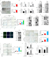
Fig. 6. LATS1/2 deletion in S1PR1 knockout cells can rescue cell proliferation and reduce cell senescence.
A Immunohistochemistry staining for S1PR1, LATS1, LATS2, and YAP in human ovarian cancer tissue. Scale bar, 50µm. B q-PCR for LATS1/2 silencing effect in S1PR1 knockout cells. Values represent mean ± SD of three independent experiments. One-way ANOVA; ***p < 0.001. C Cell counting assay results showing the proliferation ability of control cells, S1PR1 knockout cells, and S1PR1 knockout cells with LATS1/2 silencing (A2780 and ES-2). Cells (100,000) were seeded in 6-well plates. Counting was performed on days 1, 2, and 3 after inoculation and entailed 3 technical replicates per sample. One-way ANOVA; **p < 0.01, ***p < 0.001. D β-galactosidase staining results showing senescence of wild-type ovarian cancer cells, S1PR1 knockout cells, and S1PR1 knockout cells with LATS1/2 silencing. Scale bar, 50µm. Values represent mean ± SD of three independent experiments. One-way ANOVA; **p < 0.01, ***p < 0.001. E Western blot results for LATS1, LATS2, S1PR1, AKT, PI3K, PDK1, P21, P27, and P62 in wild-type ovarian cancer cells, S1PR1 knockout cells, and S1PR1 knockout cells with LATS1/2 silencing. GAPDH was used as control. F q-PCR of transcript levels of P21 in wild-type cells, S1PR1 knockout cells, and S1PR1 knockout cells with silenced P21. Values represent mean ± SD of three independent experiments. One-way ANOVA; *p < 0.05, ***p < 0.001. G β-galactosidase staining was used to analyze senescence changes in S1PR1 knockout cells with P21 silencing qualitatively (left) and quantitatively (right). Scale bar, 50µm. Values represent mean ± SD of three independent experiments. One-way ANOVA; ***p < 0.001. H Immunohistochemistry staining of S1PR1 and YAP in the liver of MST2 knockout mice. Scale bar, 50µm. Student’s t-test; ***p < 0.001. I ChIP analysis of the binding of YAP to S1PR1 promoter in ovarian cancer cells. Student’s t-test; **p < 0.01. J Schematic diagram of the S1PR1 signaling cascade. After the activation of S1PR1 in ovarian cells, the expression of PDK1 increases, which inhibits the expression of MST1/2-LATS1/2 (the key proteins in the Hippo signaling pathway) and increases YAP transcriptional activity. YAP can further promote S1PR1 transcription and tumor cell proliferation. After knocking out or inhibiting the expression of S1PR1, the expression of PI3K and PDK1 decreases, and MST1/2-LATS1/2 is activated, in turn leading to the inactivation of YAP (by phosphorylation) and tumor cell senescence.

References

    1. Lisio MA, Fu L, Goyeneche A, Gao ZH, Telleria C. High-grade serous ovarian cancer: basic sciences, clinical and therapeutic standpoints. Int J Mol Sci. 2019;20:952. doi: 10.3390/ijms20040952. - DOI - PMC - PubMed
    1. Shen L, Xia M, Zhang Y, Luo H, Dong D, Sun L. Mitochondrial integration and ovarian cancer chemotherapy resistance. Exp Cell Res. 2021;401:112549. doi: 10.1016/j.yexcr.2021.112549. - DOI - PubMed
    1. Keyvani V, Farshchian M, Esmaeili SA, Yari H, Moghbeli M, Nezhad SK, et al. Ovarian cancer stem cells and targeted therapy. J Ovarian Res. 2019;12:120. doi: 10.1186/s13048-019-0588-z. - DOI - PMC - PubMed
    1. Campisi J. Aging, cellular senescence, and cancer. Annu Rev Physiol. 2013;75:685–705. doi: 10.1146/annurev-physiol-030212-183653. - DOI - PMC - PubMed
    1. Wang Z, Liu H, Xu C. Cellular senescence in the treatment of ovarian cancer. Int J Gynecol Cancer. 2018;28:895–902. doi: 10.1097/IGC.0000000000001257. - DOI - PMC - PubMed

MeSH terms