. 2024 Mar;20(3):365-372.
doi: 10.1038/s41589-023-01434-y. Epub 2023 Oct 12.

Activation of human STING by a molecular glue-like compound

Affiliations

Activation of human STING by a molecular glue-like compound

Jie Li et al. Nat Chem Biol. 2024 Mar.

Abstract

Stimulator of interferon genes (STING) is a dimeric transmembrane adapter protein that plays a key role in the human innate immune response to infection and has been therapeutically exploited for its antitumor activity. The activation of STING requires its high-order oligomerization, which could be induced by binding of the endogenous ligand, cGAMP, to the cytosolic ligand-binding domain. Here we report the discovery through functional screens of a class of compounds, named NVS-STGs, that activate human STING. Our cryo-EM structures show that NVS-STG2 induces the high-order oligomerization of human STING by binding to a pocket between the transmembrane domains of the neighboring STING dimers, effectively acting as a molecular glue. Our functional assays showed that NVS-STG2 could elicit potent STING-mediated immune responses in cells and antitumor activities in animal models.


Conflict of interest statement

All authors (except otherwise noted) are or were at the time of their involvement with the research employees of Novartis Institute for BioMedical Research and may hold stock in Novartis. S.M.M. and K.E.S. were at the time of their involvement with the research employees of Aduro Biotech and may hold stock in Aduro (now Chinook Therapeutics).

Figures

Fig. 1
Fig. 1. Identification of NVS-STG2 as a potent allosteric small molecule STING agonist.
a, High-throughput screen of a 250,000 small-molecule compound library with the THP1-Dual reporter cell line identified NVS-STG1 (red circle). One representative from biological duplicates is shown. b, Structures of NVS-STG1 and its more potent analog NVS-STG2. AC50, half-maximal activity concentration; Amax, maximum activity. c, NVS-STG2 induces STING-dependent IRF3 phosphorylation in THP1 cells. One representative from three biological replicates is shown. d, Structures of NVS-STG3 and PAL probe, NVS-STG4. e, Identification of proteins that are enriched by NVS-STG4 and where this labeling can be competed by NVS-STG3. The x axis denotes the enrichment observed in the presence of 1 µM NVS-STG4 relative to 0 µM NVS-STG4 (log2(0 µM NVS-STG4/1 µM NVS-STG4)). STING(TMEM173) was enriched over twofold with PAL probe NVS-STG4 over no probe control in chemoproteomics pull-down. Proteins below the dashed horizontal line show reduction in the enrichment in the presence of 20 µM NVS-STG3. STING(TMEM173) was also competed by adding NVS-STG3. f, NVS-STG2 selectively activates hSTING (solid circle), but not mSTING (solid square). A fusion STING with hSTING N-terminal TMD (1–153) and mouse C-terminal LBD (153–378) responds to NVS-STG2 (empty circle). g, hSTING mutants (S27V, V31M, L93I, R95C, I103S, P115I) were generated according to key amino acid differences between human and mouse STING N-terminal TMD. NVS-STG2 activity is sensitive to hSTING R95C mutation. h, Comparison of residues in human and mouse STING tested by mutation in g. In f and g, the axis represents concentrations of STG2 in log scale, and the symbols and error bars represent mean and s.e.m., respectively, from three biological replicates. KO, knockout; MW, molecular weight. Source data
Fig. 2
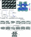
Fig. 2. Cryo-EM structures of hSTING bound to NVS-STG2.
a, Induction of STING oligomerization by cGAMP, C53, NVS-STG2 and their different combinations. STG2, NVS-STG2. One representative gel from three replicates is shown. b, Micrographs of STING bound to cGAMP/NVS-STG2 or cGAMP/NVS-STG2/C53. Images shown are representatives from n = 3,352 and n = 4,921 images for the cGAMP/NVS-STG2 and cGAMP/NVS-STG2/C53 samples, respectively. Arrows highlight STING oligomers. c, Cryo-EM density map and atomic model of STING bound to cGAMP/NVS-STG2. d, Cryo-EM density map and atomic model of STING bound to cGAMP/NVS-STG2/C53. TM, transmembrane. Source data
Fig. 3
Fig. 3. Details of the binding mode of NVS-STG2.
a, Overviews of the binding modes of NVS-STG2 (STG2) and C53. b, Detailed views of the NVS-STG2-binding site. c, Reponses of hSTING to NVS-STG2, with key binding pocket mutations. The x axis represents the concentration of NVS-STG2 in log scale. d, Activation of hSTING R95 mutants by STG2. In c and d, data are mean and s.e.m. from three biological replicates. Source data
Fig. 4
Fig. 4. NVS-STG2 induces antitumor activity and immune response in hSTING knock-in mice.
a, NVS-STG2 (red squares) or vehicle (black circles) was injected intratumorally to MC38 flank tumors on days 11, 14 and 18, and tumor size was measured by caliper. Data are mean ± s.e.m. (n = 9 mice in each group). b, Individual tumor growth is shown. Four of nine mice treated with NVS-STG2 (red squares) saw no tumor growth during the 33-day experimental period. c, NVS-STG2 at 400 μg (squares), 800 μg (triangles) or vehicle (circles) was injected intratumorally on day 8 to B16-SIY flank tumors and tumor size was measured by caliper. Data are mean ± s.e.m. (n = 9 mice in each group). d, NVS-STG2 induces significant IFNγ responses at 6 h after dosing by one-way analysis of variance (ANOVA), with P = 0.0008 (400 μg) and P < 0.0001 (800 μg) in Dunnett’s multiple comparisons test (mean ± s.d., n = 9 mice in each group). e, NVS-STG2 (800 μg) induces significant anti-SIY tumor T cell response at 6 days after treatment by one-way ANOVA, with P = 0.0009 in Dunnett’s multiple comparisons test (lines represent medians in scatter plots, n = 8 mice in each group). ***P < 0.001; ****P < 0.0001; NS, not significant (P > 0.05). Source data
Extended Data Fig. 1
Extended Data Fig. 1. NVS-STG2 is an allosteric small molecule human STING agonist.
a, cGAMP (blue circle, n = 14 independent samples), but not NVS-STG1 (green triangle, n = 2 independent samples), induces a dramatic (>10 °C) thermal shift in human STING C-terminal LBD (155-341) in differential scanning fluorimetry as compared to DMSO treated samples (red square, n = 14 independent samples). b, NVS-STG1-4 compound activity in comparison to cGAMP dose response curve in the activation of human STING in the THP1-Dual reporter cell line (n = 2 independent samples in each compound dose). c and d, STING LBD mutants Y240C (triangle) and R238A (square) lose response to cGAMP entirely but retain full response to NVS-STG2 (mean +/− SD, n = 3 independent samples in each compound dose). e, cGAMP activates human STING (solid circle), mouse STING (solid square), as well as a fusion STING with human STING N-terminal TMD (1-153) and mouse C-terminal LBD (153-378) (empty circle), mean +/− SD, n = 3 independent samples in each compound dose. f, Mouse LBD selective ligand DMXAA activates mouse STING (solid square), as well as a fusion STING with human STING N-terminal TMD (1-153) and mouse C-terminal LBD (153-378) (empty circle), but not human STING (solid circle), mean +/− SD, n = 3 independent samples in each compound dose. g, Mouse STING C95R mutant was not sufficient to render human STING selective NVS-STG2 active toward mouse STING (mean +/− SD, n = 3 independent samples in each compound dose). Source data
Extended Data Fig. 2
Extended Data Fig. 2. Image processing procedure of STING bound to cGAMP/NVS-STG2.
a, 2D class averages of segmented high-order oligomers of STING. b, Local resolution map of the final 3D reconstruction. c, Gold-standard FSC curve of the final 3D reconstruction (left) and FSC between the map and atomic model (right). d, Image processing procedure.
Extended Data Fig. 3
Extended Data Fig. 3. Image processing procedure of STING bound to cGAMP/NVS-STG2/C53.
a, 2D class averages of segmented high-order oligomers of STING. b, Local resolution map of the final 3D reconstruction. c, Gold-standard FSC curve of the final 3D reconstruction (left) and FSC between the map and atomic model (right). d, Image processing procedure.
Extended Data Fig. 4
Extended Data Fig. 4. Sample density maps of various parts of the structure.
Various parts of the structure of STING bound to cGAMP/NVS-STG2/C53 are show as sticks with the cryo-EM density superimposed.
Extended Data Fig. 5
Extended Data Fig. 5. Comparison of STING bound to different ligands.
a and b, Superimposition of the STING tetramer bound to cGAMP/NVS-STG2/C53, cGAMP/STG2 or cGAMP/C53 (PDB ID: 7SII). The three structures have high similarity to one another. c, NVS-STG2 induces a small conformational change around L136.
Extended Data Fig. 6
Extended Data Fig. 6. STG2-induced oligomerization of STING is dependent on R95.
a, Induction of oligomerization of wild-type (WT) STING and the R95A mutant by cGAMP, NVS-STG2 or their combination. STG2, NVS-STG2. b, Fluorescent puncta-like structure formation of wild-type STING and the R95A mutant upon activation by cGAMP or NVS-STG2 (scale bar 20 µm). Data shown are representatives of three biological replicates. Source data
Extended Data Fig. 7
Extended Data Fig. 7. Characterization of NVS-STG2 in human PBMC and human STING KI mice BMDM.
a, NVS-STG2 induces high level of IFN-b in all major human PBMC haplotypes (WT, REF and HAQ), in comparable level to endogenous ligand cGAMP (mean fold changes over untreated sample +/− SEM, n = 3 independent samples for each condition). b, Diagram of human STING CRISPR KI. c, Bone marrow derived macrophages (BMDM) from hSTING knock-in (KI) mice (mSTINGwt/wt, mSTINGindel/indel, hSTING+/indel, hSTING+/+) were stimulated with cGAMP (green) or NVS-STG2(red) and IP10 levels (n = 1 for each condition) were measured. cGAMP induces IP10 in both mouse and human STING mice BMDM, while NVS-STG2 only induces IP10 in mice with hSTING KI alleles. Source data

Comment in

  • Molecular glue-like STING activator.
    Crunkhorn S. Crunkhorn S. Nat Rev Drug Discov. 2023 Dec;22(12):955. doi: 10.1038/d41573-023-00173-y. Nat Rev Drug Discov. 2023. PMID: 37907755 No abstract available.

References

    1. Ishikawa H, Barber GN. STING is an endoplasmic reticulum adaptor that facilitates innate immune signalling. Nature. 2008;455:674–678. doi: 10.1038/nature07317. - DOI - PMC - PubMed
    1. Zhang X, Bai XC, Chen ZJ. Structures and mechanisms in the cGAS-STING innate immunity pathway. Immunity. 2020;53:43–53. doi: 10.1016/j.immuni.2020.05.013. - DOI - PubMed
    1. Zhang H, You QD, Xu XL. Targeting stimulator of interferon genes (STING): a medicinal chemistry perspective. J. Med. Chem. 2020;63:3785–3816. doi: 10.1021/acs.jmedchem.9b01039. - DOI - PubMed
    1. Le Naour J, Zitvogel L, Galluzzi L, Vacchelli E, Kroemer G. Trial watch: STING agonists in cancer therapy. Oncoimmunology. 2020;9:1777624. doi: 10.1080/2162402X.2020.1777624. - DOI - PMC - PubMed
    1. Ding, C., Song, Z., Shen, A., Chen, T. & Zhang, A. Small molecules targeting the innate immune cGAS-STING-TBK1 signaling pathway. Acta Pharm. Sin. B10, 2272–2298 (2020). - PMC - PubMed

Publication types