

Application and prospects of AI-based radiomics in ultrasound diagnosis
- PMID: 37828411
- PMCID: PMC10570254
- DOI: 10.1186/s42492-023-00147-2
Application and prospects of AI-based radiomics in ultrasound diagnosis
Abstract
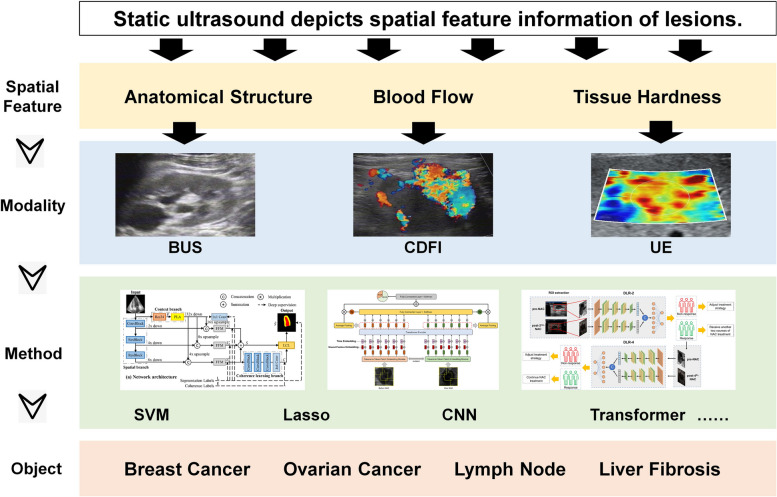
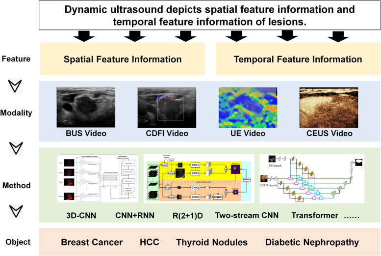
Artificial intelligence (AI)-based radiomics has attracted considerable research attention in the field of medical imaging, including ultrasound diagnosis. Ultrasound imaging has unique advantages such as high temporal resolution, low cost, and no radiation exposure. This renders it a preferred imaging modality for several clinical scenarios. This review includes a detailed introduction to imaging modalities, including Brightness-mode ultrasound, color Doppler flow imaging, ultrasound elastography, contrast-enhanced ultrasound, and multi-modal fusion analysis. It provides an overview of the current status and prospects of AI-based radiomics in ultrasound diagnosis, highlighting the application of AI-based radiomics to static ultrasound images, dynamic ultrasound videos, and multi-modal ultrasound fusion analysis.
Keywords: Artificial intelligence; B-mode ultrasound; Color Doppler flow imaging; Contrast-enhanced ultrasound; Deep learning; Multimodal ultrasound; Radiomics; Ultrasound elastography; Ultrasound imaging.
© 2023. China Graphics Society.
Conflict of interest statement
The authors declare no conflict of interest.
Figures

Similar articles
-
AI-Based Radiological Imaging for HCC: Current Status and Future of Ultrasound.Diagnostics (Basel). 2021 Feb 12;11(2):292. doi: 10.3390/diagnostics11020292. Diagnostics (Basel). 2021. PMID: 33673229 Free PMC article. Review.
-
Preoperative diagnosis and prediction of hepatocellular carcinoma: Radiomics analysis based on multi-modal ultrasound images.BMC Cancer. 2018 Nov 12;18(1):1089. doi: 10.1186/s12885-018-5003-4. BMC Cancer. 2018. PMID: 30419849 Free PMC article.
-
Artificial intelligence - based ultrasound elastography for disease evaluation - a narrative review.Front Oncol. 2023 Jun 2;13:1197447. doi: 10.3389/fonc.2023.1197447. eCollection 2023. Front Oncol. 2023. PMID: 37333814 Free PMC article. Review.
-
Articles That Use Artificial Intelligence for Ultrasound: A Reader's Guide.Front Oncol. 2021 Jun 10;11:631813. doi: 10.3389/fonc.2021.631813. eCollection 2021. Front Oncol. 2021. PMID: 34178622 Free PMC article. Review.
-
Advances and prospects of multi-modal ophthalmic artificial intelligence based on deep learning: a review.Eye Vis (Lond). 2024 Oct 1;11(1):38. doi: 10.1186/s40662-024-00405-1. Eye Vis (Lond). 2024. PMID: 39350240 Free PMC article. Review.
Cited by
-
Ultrasound-based radiogenomics: status, applications, and future direction.Ultrasonography. 2025 Mar;44(2):95-111. doi: 10.14366/usg.24152. Epub 2024 Dec 12. Ultrasonography. 2025. PMID: 39935290 Free PMC article.
-
Ultrasomics differentiation of malignant and benign focal liver lesions based on contrast-enhanced ultrasound.BMC Med Imaging. 2024 Sep 16;24(1):242. doi: 10.1186/s12880-024-01426-x. BMC Med Imaging. 2024. PMID: 39285357 Free PMC article.
-
Artificial intelligence-assisted diagnosis of early allograft dysfunction based on ultrasound image and data.Vis Comput Ind Biomed Art. 2025 May 12;8(1):13. doi: 10.1186/s42492-025-00192-z. Vis Comput Ind Biomed Art. 2025. PMID: 40353942 Free PMC article.
-
Assessing the efficacy of ultrasound deep tissue evaluation in predicting hospital-acquired pressure injuries: A diagnostic study.Medicine (Baltimore). 2025 Jul 11;104(28):e43268. doi: 10.1097/MD.0000000000043268. Medicine (Baltimore). 2025. PMID: 40660566 Free PMC article.
-
Multi-task approach based on combined CNN-transformer for efficient segmentation and classification of breast tumors in ultrasound images.Vis Comput Ind Biomed Art. 2024 Jan 26;7(1):2. doi: 10.1186/s42492-024-00155-w. Vis Comput Ind Biomed Art. 2024. PMID: 38273164 Free PMC article.
References
-
- Ma LF, Wang R, He Q, Huang LJ, Wei XY, Lu X et al (2022) Artificial intelligence-based ultrasound imaging technologies for hepatic diseases. iLIVER 1(4):252–264. 10.1016/j.iliver.2022.11.001
-
- Janiesch C, Zschech P, Heinrich K. Machine learning and deep learning. Electron Markets. 2021;31(3):685–695. doi: 10.1007/s12525-021-00475-2. - DOI
Publication types
LinkOut - more resources
Full Text Sources