. 2023 Sep 18;14(39):10962-10970.
doi: 10.1039/d3sc03822j. eCollection 2023 Oct 11.

Post-synthetic benzylation of the mRNA 5' cap via enzymatic cascade reactions

Affiliations

Post-synthetic benzylation of the mRNA 5' cap via enzymatic cascade reactions

N V Cornelissen et al. Chem Sci. .

Abstract

mRNAs are emerging modalities for vaccination and protein replacement therapy. Increasing the amount of protein produced by stabilizing the transcript or enhancing translation without eliciting a strong immune response are major steps towards overcoming the present limitations and improving their therapeutic potential. The 5' cap is a hallmark of mRNAs and non-natural modifications can alter the properties of the entire transcript selectively. Here, we developed a versatile enzymatic cascade for regioselective benzylation of various biomolecules and applied it for post-synthetic modification of mRNA at the 5' cap to demonstrate its potential. Starting from six synthetic methionine analogues bearing (hetero-)benzyl groups, S-adenosyl-l-methionine analogues are formed and utilized for N7G-cap modification of mRNAs. This post-synthetic enzymatic modification exclusively modifies mRNAs at the terminal N7G, producing mRNAs with functional 5' caps. It avoids the wrong orientation of the 5' cap-a problem in common co-transcriptional capping. In the case of the 4-chlorobenzyl group, protein production was increased to 139% during in vitro translation and to 128-150% in four different cell lines. This 5' cap modification did not activate cytosolic pathogen recognition receptors TLR3, TLR7 or TLR8 significantly more than control mRNAs, underlining its potential to contribute to the development of future mRNA therapeutics.


Conflict of interest statement

There are no conflicts to declare.

Figures

Fig. 1
Fig. 1. Cascade reactions for methylation in nature and application for site-specific alkylation and benzylation. (A) Methionine and ATP are reacted by methionine adenosyltransferase (MAT) to form the cofactor S-adenosyl-l-methionine (AdoMet or SAM). AdoMet can be used by methyltransferases (MTases) for the stereo-, regio- and chemo-selective methylation of DNA, RNA, proteins and small molecules. (B) Synthetic methionine analogues are used in MAT/MTase cascade reactions to transfer non-natural alkyl moieties to biomolecules. (C) Photocleavable groups derived from the 2-nitrobenzyl group can be transferred by an engineered MAT/MTase cascade to photocage DNA. (D) This work shows that MAT/MTase cascades are a general tool to transfer (hetero)-benzylic moieties and can be used to modify mRNA at the 5′ cap.
Fig. 2
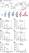
Fig. 2. MAT reaction with methionine and benzylic methionine analogues. (A) Reaction scheme with methionine 1a and benzylic methionine analogues (1b–g) prepared for this study. The AdoMet analogues (2b–g) and heat-induced degradation products (MTA analogues, 3a–g) are monitored to circumvent effects from product inhibition. PC-MjMAT is an engineered MAT variant from Methanocaldococcus jannaschii (MjMAT L147A/I351A). (B–G) HPLC analyses of reactions with the indicated amino acid performed at 37 °C or 65 °C. Conditions: 5 mM 1b–g, 5 mM ATP, 100 μM PC-MjMAT at 37 °C or 65 °C for 1 h in a total volume of 30 μL. Reaction buffer (1×) consisted of 50 mM HEPES, 10 mM MgCl2, 5 mM KCl (pH = 7.4). Adenosine (A) results from background depurination.
Fig. 3
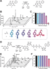
Fig. 3. Substrate scope of enzymatic benzylation of small molecules in two MAT/MTase cascade reactions. (A) The PC-MjMAT/NovO cascade converts 4,5,7-trihydroxy-3-phenylcoumarin (4) to derivatives 4a–g, benzylated at the C8-position. (B) Representative HPLC analyses of the cascade reactions of 4 with 1a–g. (C) Evaluation of conversions from 4 to 4a–g calculated from the peak area (n = 3). Conditions: 5 mM 1a–g, 5 mM ATP, 100 μM PC-MjMAT, 50 μM NovO, 10 μM MTAN, 1 mM 4 at 37 °C for 2 h. (D) The PC-MjMAT/Ecm1 cascade converts the dinucleotide GpppA (5) to derivatives 5a–g, benzylated at the N7-position of guanosine. (E) Representative HPLC analyses of the cascade reactions of 5 with 1a–g. (F) Evaluation of conversions from 5 to 5a–g calculated from peak area (n = 3). Conditions: 5 mM 1a–g, 5 mM ATP, 100 μM PC-MjMAT, 50 μM Ecm1, 10 μM MTAN, 500 μM 5 at 37 °C for 2 h.
Fig. 4
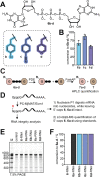
Fig. 4. Enzymatic generation of N7-modified GpppG caps and analysis of post-transcriptionally modified GpppG-RNAs. (A) Structure of N7-modified GpppG caps (6b–d). (B) Conversion of GpppG to 6b–d calculated from the HPLC peak area (n = 3). Conditions: 5 mM 1b/c or 8 mM 1d, 5 mM ATP, 100 μM PC-MjMAT, 50 μM Ecm1, 10 μM MTAN, 2 mM 6 at 37 °C for 2 h. (C) Determination of the concentration of purified 5′ caps 6b–d. 6b–d were digested into nucleosides using snake venom phosphodiesterase (PDE), dephosphorylated using FastAP and quantified by HPLC using an external standard. (D) Scheme illustrating post-synthetic modification to obtain mRNAs with different 5′ cap modifications. GpppG-mRNAs are modified via the enzymatic PC-MjMAT/Ecm1 cascade. Nuclease P1 digests the mRNA while leaving the 5′ caps (6, 6a–d) intact for LC-QqQ analysis. (E) Analysis of mRNA integrity after the PC-MjMAT/Ecm1 cascade. M: marker, control: GpppG-RNA, 6-RNA: GpppG-RNA incubated in PC-MjMAT/Ecm1 reaction mixture without enzymes. Reaction conditions: 0.2 μM mRNA, 2.5 μM PC-MjMAT, 2.5 μM MTAN, 50 μM 1a–d, 50 μM ATP, 1 μM Ecm1 in 0.5× NEB CutSmart buffer at 37 °C for 3 h. (F) Conversion of post-synthetic 5′ cap modification of GpppG-mRNA via the chemo-enzymatic cascade. Results show LC-QqQ-MS analysis, as illustrated in (D).
Fig. 5
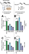
Fig. 5. Effect of mRNA 5′ cap methylation and benzylation on the amount of protein produced in vitro and in cells. Co- and post-transcriptional 5′ cap modification strategies were evaluated in parallel. (A and C) Luciferase activity from differently capped RLuc-mRNAs prepared via in vitro transcription (IVT) and translated in rabbit reticulocyte lysate (RRL) in vitro (A) or in HeLa cells (C). (B and D) Same assays, but with RLuc-mRNAs modified post-transcriptionally via the PC-MjMAT/Ecm1 cascade. The results are normalized to ARCA-RLuc-mRNA. The average value ± SD of n = 3 independent experiments is shown. Statistical analysis: unpaired t-test. p < 0.05: *, p < 0.01: **, p < 0.001: ***, n.s.: not significant.
Fig. 6
Fig. 6. (A) Scheme of HPLC-based 5′ cap orientation assay. m26a–d (terminal = correct orientation), 6a–d (internal = wrong orientation). (B and C) Representative HPLC analysis of the 5′ cap incorporation assay in co-transcriptionally modified RNA using 6a–b, respectively. (D) Evaluation of 5′ cap orientation assay for mRNAs with 6a–d (n = 3). (E) Integrity of RNA 24mer used in 5′ cap orientation assay, analysed by PAA gel (15% TBE).
Fig. 7
Fig. 7. Effect of mRNA 5′ cap methylation and benzylation on the immune response and protein production in HEK-NF-κB reporter cell lines. Co- and post-transcriptional 5′ cap modification strategies were evaluated in parallel. (A) Concept of activation of pathogen recognition receptors such as PKR, MDA5, and RIG-I in the HEK-NF-κB null cell line triggering Firefly luciferase (FLuc) expression. Created with http://BioRender.com. (B–E) Indicated HEK-NF-κB cells (Null, TLR3, TLR7, TLR8) were transfected with differently capped RLuc-mRNAs and NF-κB activation was quantified via FLuc activity. Data are normalized to the ARCA-mRNA. (F–I) RLuc activity as a measure of protein production in indicated HEK-NF-κB cells transfected with differently capped RLuc-mRNAs. Data of n = 3 independent experiments are shown as average values ± SD. Statistical analysis: unpaired t-test. p < 0.05: *, p < 0.01: **, p < 0.001: ***, n.s.: not significant.

References

    1. Jackson L. A. Anderson E. J. Rouphael N. G. Roberts P. C. Makhene M. Coler R. N. McCullough M. P. Chappell J. D. Denison M. R. Stevens L. J. Pruijssers A. J. McDermott A. Flach B. Doria-Rose N. A. Corbett K. S. Morabito K. M. O'Dell S. Schmidt S. D. Swanson 2nd P. A. Padilla M. Mascola J. R. Neuzil K. M. Bennett H. Sun W. Peters E. Makowski M. Albert J. Cross K. Buchanan W. Pikaart-Tautges R. Ledgerwood J. E. Graham B. S. Beigel J. H. N. Engl. J. Med. 2020;383:1920–1931. doi: 10.1056/NEJMoa2022483. - DOI - PMC - PubMed
    1. Lorentzen C. L. Haanen J. B. Met Ö. Svane I. M. Lancet Oncol. 2022;23:e450–e458. doi: 10.1016/S1470-2045(22)00372-2. - DOI - PMC - PubMed
    1. Ramaswamy S. Tonnu N. Tachikawa K. Limphong P. Vega J. B. Karmali P. P. Chivukula P. Verma I. M. Proc. Natl. Acad. Sci. U. S. A. 2017;114:E1941–e1950. doi: 10.1073/pnas.1619653114. - DOI - PMC - PubMed
    2. Perez-Garcia C. G. Diaz-Trelles R. Vega J. B. Bao Y. Sablad M. Limphong P. Chikamatsu S. Yu H. Taylor W. Karmali P. P. Tachikawa K. Chivukula P. Mol. Ther.--Nucleic Acids. 2022;28:87–98. doi: 10.1016/j.omtn.2022.02.020. - DOI - PMC - PubMed
    1. Rohner E. Yang R. Foo K. S. Goedel A. Chien K. R. Nat. Biotechnol. 2022;40:1586–1600. doi: 10.1038/s41587-022-01491-z. - DOI - PubMed
    1. Strzelecka D. Smietanski M. Sikorski P. J. Warminski M. Kowalska J. Jemielity J. RNA. 2020;26:1815–1837. doi: 10.1261/rna.077099.120. - DOI - PMC - PubMed
    2. Wojtczak B. A. Sikorski P. J. Fac-Dabrowska K. Nowicka A. Warminski M. Kubacka D. Nowak E. Nowotny M. Kowalska J. Jemielity J. J. Am. Chem. Soc. 2018;140:5987–5999. doi: 10.1021/jacs.8b02597. - DOI - PubMed
    3. Drazkowska K. Tomecki R. Warminski M. Baran N. Cysewski D. Depaix A. Kasprzyk R. Kowalska J. Jemielity J. Sikorski P. J. Nucleic Acids Res. 2022;50:9051–9071. doi: 10.1093/nar/gkac722. - DOI - PMC - PubMed