Case Reports
. 2023 Sep 27:13:1263556.
doi: 10.3389/fonc.2023.1263556. eCollection 2023.

Memory deficit following resection of an intraventricular myxoid glioneuronal tumor impinging on the bilateral fornix: A case report

Affiliations
Case Reports

Memory deficit following resection of an intraventricular myxoid glioneuronal tumor impinging on the bilateral fornix: A case report

Alena Stasenko et al. Front Oncol. .

Abstract

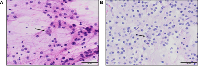
Background: Recently recognized as a distinct entity, a myxoid glioneuronal tumor (MGNT) is a rare, low-grade central nervous system tumor. MGNTs are commonly located at the septum pellucidum or in the third ventricle, increasing the likelihood of tumor or treatment-related damage to adjacent structures critical for memory, such as the fornix. Though there have been a handful of case reports of neurosurgical and oncological outcomes of MGNTs, memory outcomes following resection of MGNTs adjacent to the fornix have not been previously reported.

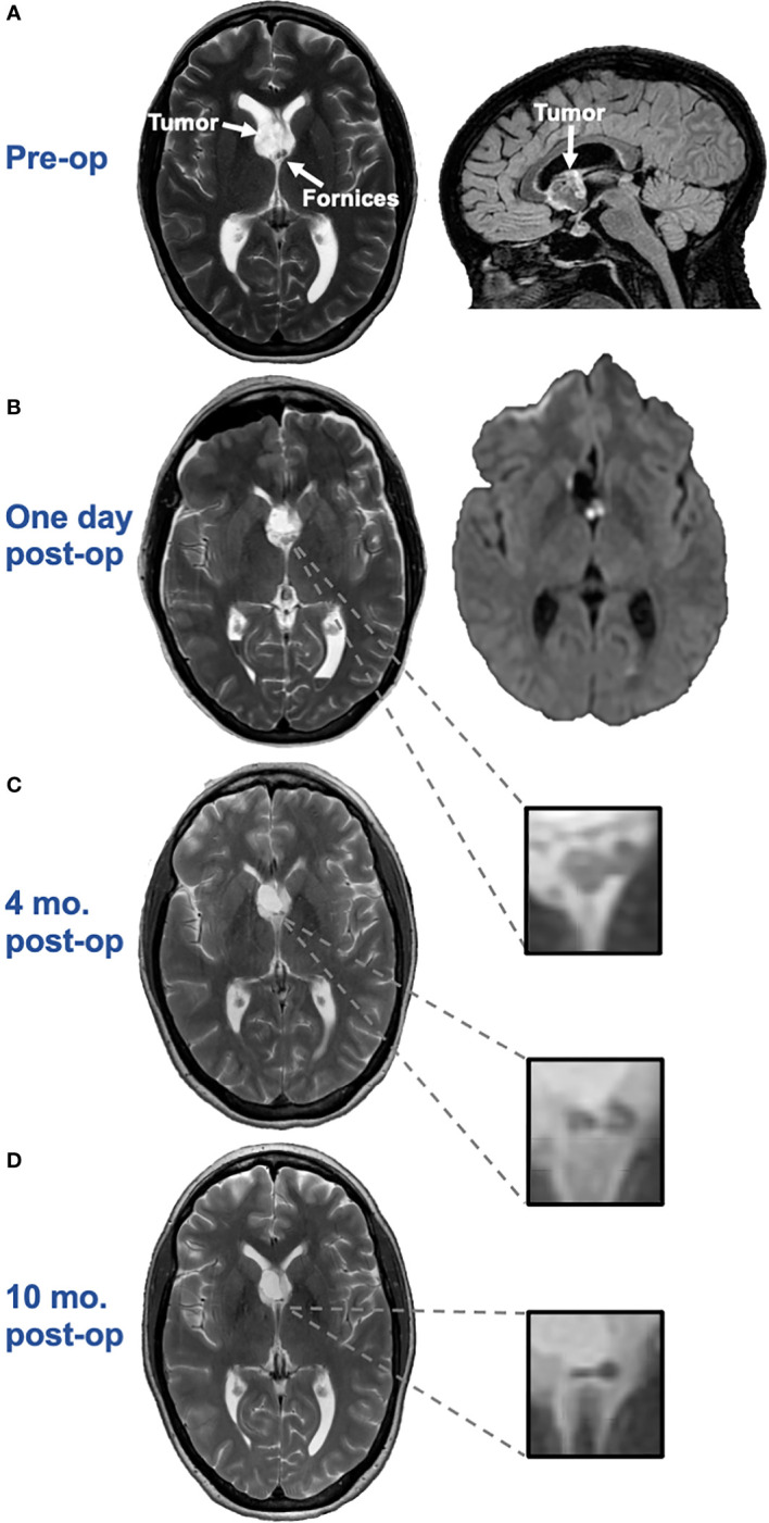
Methods: We present a case of a high functioning female for whom an MRI revealed an incidental finding of an intraventricular tumor adjacent to the fornix bilaterally. The patient underwent resection of the tumor followed by MRI surveillance without additional oncologic intervention. Due to reported cognitive problems, the patient was referred for serial neuropsychological evaluations.

Results: Post-operative MRI following resection revealed cytotoxic edema followed by selective, progressive atrophy of the bilateral anterior fornices. Post-surgically, the patient developed an isolated verbal memory impairment, which persisted one-year post resection with minimal improvement. The memory impairment impacted the patient's everyday functioning, including the ability to work in a cognitively demanding job.

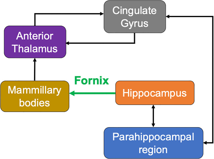
Conclusion: This unique case demonstrates the critical role of the bilateral fornix in verbal memory and underscores the importance of a careful risk/benefit analysis when considering neurosurgical intervention to MGNTs and other intracranial lesions adjacent to this structure during neurosurgical planning.

Keywords: fornix; intraventricular; memory; myxoid glioneuronal tumor; neurosurgery; resection.


Conflict of interest statement

The authors declare that the research was conducted in the absence of any commercial or financial relationships that could be construed as a potential conflict of interest.

Figures

Figure 1
Figure 1
(A). Axial T2 (left) and sagittal 3D CUBE FLAIR (right) images from brain MRI approximately two months before surgery demonstrate an intraventricular mass with a bubbly appearance centered at the right aspect of the septum pellucidum, near the fornices. (B). Axial T2 image (left) and axial diffusion-weighted image (DWI) (right) one day post-surgery demonstrates swelling and restricted diffusion of the bilateral anterior fornices suggesting forniceal injury. The hyperintense signal on the axial DWI had corresponding hypointense signal on the apparent diffusion coefficient (ADC) map, not pictured here, confirming true restricted diffusion. (C). Axial T2 image approximately 4-months after surgery demonstrates irregular and mildly atrophic appearance of the fornices, compatible with evolution of previously seen cytotoxic edema in this region. (D). Axial T2 image approximately 10-months after surgery demonstrates progressive atrophic appearance of the fornices.
Figure 2
Figure 2
(A). Hematoxylin and eosin (H&E) stained squash preparation and (B). tissue section both show a dual cell population, with a predominance of uniform small round oligodendrocyte-like cells in a fibrillar background, focally accompanied by floating neurons (arrows), which appear larger, with more cytoplasm, and prominent nucleoli.
Figure 3
Figure 3
Schematic of the classic Papez circuit—a medial limbic network important for episodic memory. Information from cortical association areas is sent to the hippocampus via the cingulate gyrus, parahippocampal gyrus/entorhinal cortex, and cingulum bundle (white matter fibers). Processed information from the hippocampus is sent via the fornix (green) to the mammillary bodies of the hypothalamus. Information from the mammillary bodies is sent to the anterior nuclei of the thalamus via the mammillothalamic tract, which then projects fibers to the cingulate gyrus, parahippocampal gyrus/entorhinal cortex, and cingulum back to the hippocampus.

References

    1. Solomon DA, Korshunov A, Sill M, Jones DTW, Kool M, Pfister SM, et al. Myxoid glioneuronal tumor of the septum pellucidum and lateral ventricle is defined by a recurrent PDGFRA p.K385 mutation and DNT-like methylation profile. Acta Neuropathol (2018) 136(2):339–43. doi: 10.1007/s00401-018-1883-2 - DOI - PMC - PubMed
    1. Louis DN, Perry A, Wesseling P, Brat DJ, Cree IA, Figarella-Branger D, et al. The 2021 WHO classification of tumors of the central nervous system: A summary. Neuro Oncol (2021) 23(8):1231–51. doi: 10.1093/neuonc/noab106 - DOI - PMC - PubMed
    1. Caporalini C, Scagnet M, Giunti L, Cetica V, Mei D, Conti V, et al. Myxoid glioneuronal tumor: Histopathologic, neuroradiologic, and molecular features in a single center series. Neoplasia (2023) 37:100885. doi: 10.1016/j.neo.2023.100885 - DOI - PMC - PubMed
    1. Lucas CHG, Villanueva-Meyer JE, Whipple N, Oberheim Bush NA, Cooney T, Chang S, et al. Myxoid glioneuronal tumor, PDGFRA p.K385-mutant: clinical, radiologic, and histopathologic features. Brain Pathol (2020) 30(3):479–94. doi: 10.1111/bpa.12797 - DOI - PMC - PubMed
    1. Benear SL, Ngo CT, Olson IR. Dissecting the fornix in basic memory processes and neuropsychiatric disease: A review. Brain Connect (2020) 10(7):331–54. doi: 10.1089/brain.2020.0749 - DOI - PMC - PubMed

Publication types