

Pancreatic tail cancer in the setting of pancreatitis with a review of the literature: A case report
- PMID: 37830064
- PMCID: PMC10565090
- DOI: 10.1002/ccr3.8023
Pancreatic tail cancer in the setting of pancreatitis with a review of the literature: A case report
Abstract
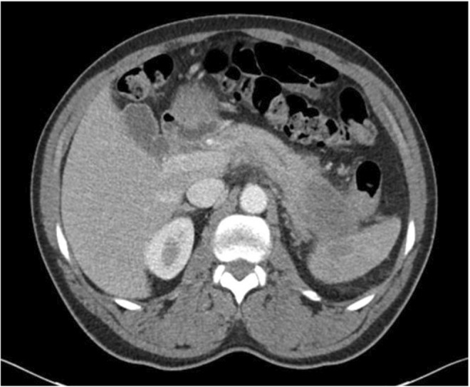
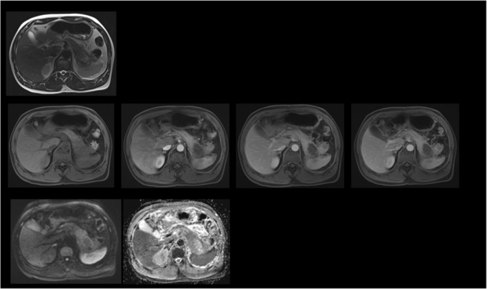
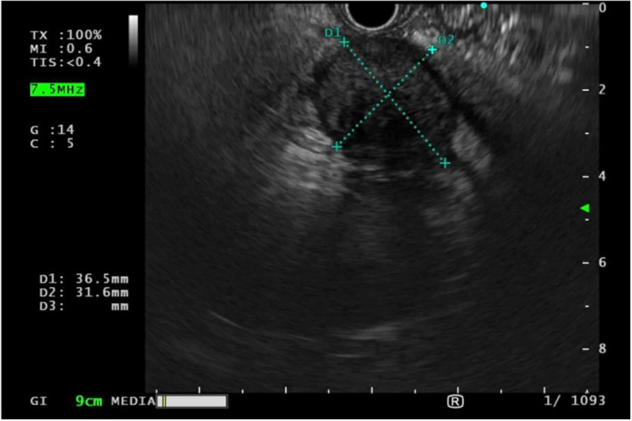
Environmental risk factors for pancreatic cancer include acute and chronic pancreatitis, obesity, and tobacco use. Differentiating a pancreatic neoplasm in a patient with pancreatitis can be challenging due to their similar presentations. A 57-year-old African American man with a history of congestive heart failure, pancreatitis, and incomplete pancreas divisum presented with an epigastric abdominal pain that radiated to his back. Imaging showed necrotizing pancreatitis, a developing splenic infarct, and a mass at the pancreas tail. The patient was discharged with pain medications and was recommended follow-up imaging after resolution of his pancreatitis. He was readmitted to the emergency department 2 weeks later with recurrent acute abdominal pain. Computed tomography scan of abdomen and pelvis followed by magnetic resonance imaging and endoscopic ultrasound revealed an infiltrative pancreatic tail mass. Biopsy of the mass confirmed a locally advanced pancreatic tail adenocarcinoma. Chronic pancreatitis is associated with pancreatic cancer. Practitioners should be aware of the co-existence of chronic pancreatitis and pancreatic cancer, and the initial steps to evaluate a malignancy in chronic pancreatitis.
Keywords: chronic pancreatitis; imaging; pancreatic ductal adenocarcinoma; pancreatic neoplasm.
© 2023 The Authors. Clinical Case Reports published by John Wiley & Sons Ltd.
Conflict of interest statement
The authors declare that they have no conflicts of interest.
Figures
Similar articles
-
The seemingly innocuous presentation of metastatic pancreatic tail cancer: a case report.J Med Case Rep. 2019 Jun 11;13(1):178. doi: 10.1186/s13256-019-2125-5. J Med Case Rep. 2019. PMID: 31182138 Free PMC article.
-
Pancreas divisum causing recurrent pancreatitis in a young patient: A case report.Radiol Case Rep. 2023 Jul 28;18(10):3535-3538. doi: 10.1016/j.radcr.2023.07.028. eCollection 2023 Oct. Radiol Case Rep. 2023. PMID: 37547796 Free PMC article.
-
Acute pancreatitis: pancreas divisum with ventral duct intraductal papillary mucinous neoplasms.BMJ Case Rep. 2014 Oct 7;2014:bcr2014205322. doi: 10.1136/bcr-2014-205322. BMJ Case Rep. 2014. PMID: 25293684 Free PMC article.
-
Neuroendocrine carcinoma of the minor papilla with pancreas divisum: a case report and review of the literature.J Med Case Rep. 2023 Mar 28;17(1):127. doi: 10.1186/s13256-023-03828-x. J Med Case Rep. 2023. PMID: 36973791 Free PMC article.
-
Double plastic stent implantation for recurrent acute pancreatitis with incomplete pancreas divisum: a case report and literature review.J Int Med Res. 2021 Nov;49(11):3000605211060142. doi: 10.1177/03000605211060142. J Int Med Res. 2021. PMID: 34842463 Free PMC article. Review.
References
-
- Kirkegård J, Cronin‐Fenton D, Heide‐Jørgensen U, Mortensen FV. Acute pancreatitis and pancreatic cancer risk: a Nationwide Matched‐Cohort Study in Denmark. Gastroenterology. 2018;154:1729‐1736. - PubMed
-
- Lowenfels AB, Maisonneuve P, Cavallini G, et al. Pancreatitis and the risk of pancreatic cancer. N Engl J Med. 1993;328:1433‐1437. - PubMed
-
- Munigala S, Kanwal F, Xian H, Scherrer JF, Agarwal B. Increased risk of pancreatic adenocarcinoma after acute pancreatitis. Clin Gastroenterol Hepatol. 2014;12:1143‐1150.e1. - PubMed
Publication types
LinkOut - more resources
Full Text Sources
