. 2024 Jan;30(1):e14486.
doi: 10.1111/cns.14486. Epub 2023 Oct 13.

Dexmedetomidine alleviates cognitive impairment by promoting hippocampal neurogenesis via BDNF/TrkB/CREB signaling pathway in hypoxic-ischemic neonatal rats

Affiliations

Dexmedetomidine alleviates cognitive impairment by promoting hippocampal neurogenesis via BDNF/TrkB/CREB signaling pathway in hypoxic-ischemic neonatal rats

Xiaohui Chen et al. CNS Neurosci Ther. 2024 Jan.

Abstract

Aims: Dexmedetomidine (DEX) has been reported to alleviate hypoxic-ischemic brain damage (HIBD) in neonates. This study aimed to investigate whether DEX improves cognitive impairment by promoting hippocampal neurogenesis via the BDNF/TrkB/CREB signaling pathway in neonatal rats with HIBD.

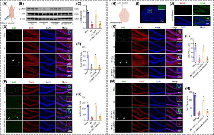
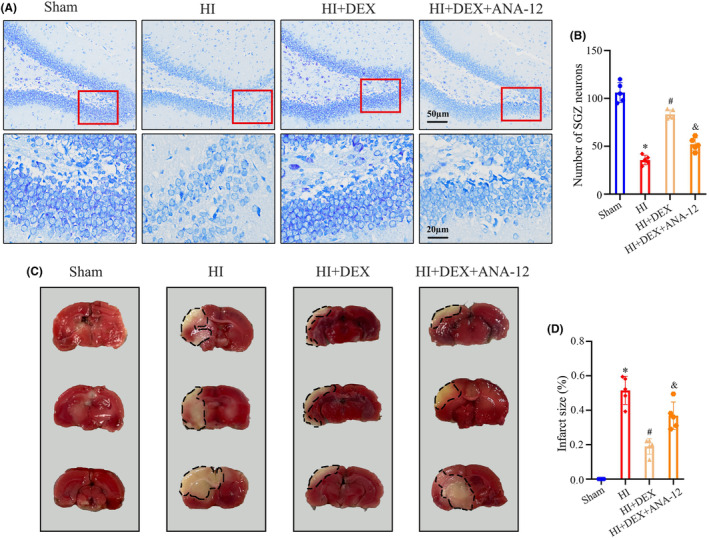
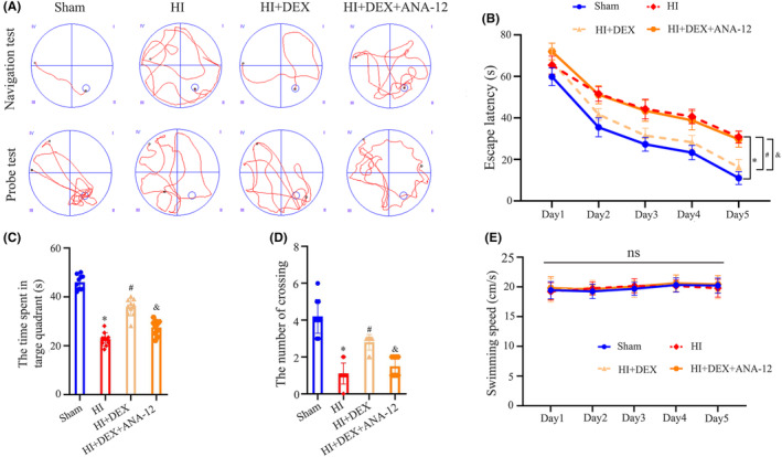
Methods: HIBD was induced in postnatal day 7 rats using the Rice-Vannucci method, and DEX (25 μg/kg) was administered intraperitoneally immediately after the HIBD induction. The BDNF/TrkB/CREB pathway was regulated by administering the TrkB receptor antagonist ANA-12 through intraperitoneal injection or by delivering adeno-associated virus (AAV)-shRNA-BDNF via intrahippocampal injection. Western blot was performed to measure the levels of BDNF, TrkB, and CREB. Immunofluorescence staining was utilized to identify the polarization of astrocytes and evaluate the levels of neurogenesis in the dentate gyrus of the hippocampus. Nissl and TTC staining were performed to evaluate the extent of neuronal damage. The MWM test was conducted to evaluate spatial learning and memory ability.

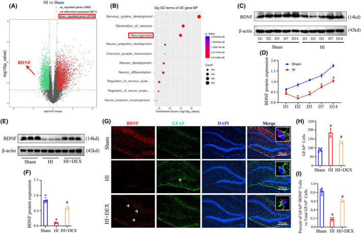
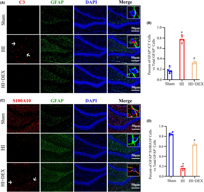
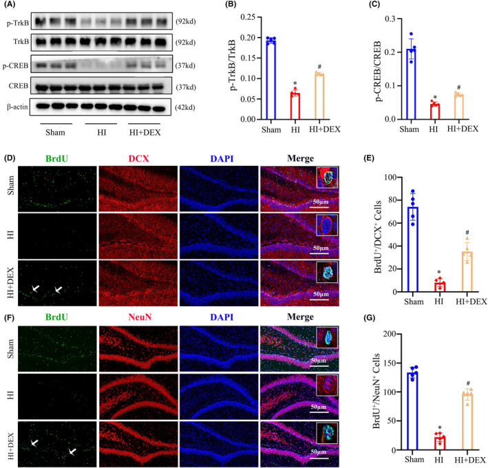
Results: The levels of BDNF and neurogenesis exhibited a notable decrease in the hippocampus of neonatal rats after HIBD, as determined by RNA-sequencing technology. Our results demonstrated that treatment with DEX effectively increased the protein expression of BDNF and the phosphorylation of TrkB and CREB, promoting neurogenesis in the dentate gyrus of the hippocampus in neonatal rats with HIBD. Specifically, DEX treatment significantly augmented the expression of BDNF in hippocampal astrocytes, while decreasing the proportion of detrimental A1 astrocytes and increasing the proportion of beneficial A2 astrocytes in neonatal rats with HIBD. Furthermore, inhibiting the BDNF/TrkB/CREB pathway using either ANA-12 or AAV-shRNA-BDNF significantly counteracted the advantageous outcomes of DEX on hippocampal neurogenesis, neuronal survival, and cognitive improvement.

Conclusions: DEX promoted neurogenesis in the hippocampus by activating the BDNF/TrkB/CREB pathway through the induction of polarization of A1 astrocytes toward A2 astrocytes, subsequently mitigating neuronal damage and cognitive impairment in neonates with HIBD.

Keywords: BDNF; astrocyte; cognitive impairment; hippocampal neurogenesis; hypoxic-ischemic brain damage.


Conflict of interest statement

The authors declare no competing interests.

Figures

FIGURE 1
FIGURE 1
DEX treatment increased the expression of BDNF in the hippocampus of neonatal rats following HIBD. (A) Volcano plots display the DEGs as red dots (upregulated) and blue dots (downregulated). (B) The top 10 downregulated DEGs are categorized into classes based on GO enrichment terms for the biological process. (C, D) Western blot analysis was performed to examine the expression levels of BDNF at 1, 2, 3, 7, 14 days after HI. (E, F) Western blot analysis was performed to examine the expression levels of BDNF at 2 days after HI insults and DEX treatment. (G) Immunofluorescent staining was performed to examine the expression levels of BDNF in astrocyte of the dentate gyrus in the hippocampus. (H) Quantification for the number of GFAP+ cells in the dentate gyrus of the hippocampus. (I) Quantification for the ratio of BDNF+/GFAP+ cells to the total number of GFAP+ cells in the dentate gyrus of the hippocampus. DEGs, differentially expressed genes; DEX, dexmedetomidine; HI, hypoxic‐ischemia; HIBD, hypoxic‐–ischemic brain damage. Data were expressed as the mean ± SD (n = 5 per group); *p < 0.05 vs. the sham group; # p < 0.05 vs. the HI group.
FIGURE 2
FIGURE 2
DEX treatment promoted polarization of astrocytes from the A1 to A2 phenotype in the hippocampus of neonatal after HIBD. (A) Dual immunofluorescence staining was performed to examine the number of A1 astrocytes labeling C3 and GFAP. (B) Quantification for the ratio of C3+/GFAP+ cells to the total number of GFAP+ cells in the dentate gyrus of the hippocampus. (C) Dual immunofluorescence staining was performed to examine the number of A2 astrocytes labeling S100A10 and GFAP. (D) Quantification for the ratio of S100A10+/GFAP+ cells to the total number of GFAP+ cells in the dentate gyrus of the hippocampus. DEX, dexmedetomidine; HI, hypoxic–ischemia; HIBD, hypoxic–ischemic brain damage. Data were expressed as the mean ± SD (n = 5 per group); # p < 0.05 vs. the HI group.
FIGURE 3
FIGURE 3
DEX treatment increased phosphorylation levels of TrkB and CREB and promoted hippocampal neurogenesis after HIBD. (A–C) Western blot analysis was performed to examine the levels of TrkB, CREB phosphorylation in the hippocampus at 2 days after HI and DEX treatment. The bar diagram represents the densitometric analyses of immunoblots. (D) Dual immunofluorescence staining was performed to examine the number of newly formed neuroblasts labeled with BrdU and DCX. (E) The quantification for the number of BrdU+/DCX+ cells in the dentate gyrus of the hippocampus. (F) Dual immunofluorescence staining was performed to examine the number of newly formed neurons labeled with BrdU and NeuN. (G) The quantification for the number of BrdU+/NeuN+ cells in the dentate gyrus of the hippocampus. DEX, dexmedetomidine; HI, hypoxic‐ischemia; HIBD, hypoxic‐–ischemic brain damage. Data were expressed as the mean ± SD (n = 5 per group); *p < 0.05 vs. the sham group; # p < 0.05 vs. the HI group.
FIGURE 4
FIGURE 4
Inhibiting the BDNF/TrkB/CREB signaling pathway reversed the ability of DEX to promote hippocampal neurogenesis after HIBD. (A) The schematic illustrates the administration of ANA‐12, a selective inhibitor of TrkB, via intraperitoneal injection. (B, C) Western blot analysis was performed to examine the phosphorylation levels of CREB in the hippocampus, and the bar diagram represents the densitometric analyses of p‐CREB. (D, K) Dual immunofluorescence staining was performed to examine the number of newly formed neuroblasts labeling BrdU and DCX. (E, L) The quantification for the number of BrdU+/DCX+ cells in the dentate gyrus of the hippocampus. (F, M) Dual immunofluorescence staining was performed to examine the number of newly formed neurons labeling BrdU and NeuN. (G, N) The quantification for the number of BrdU+/NeuN+ cells in the dentate gyrus of the hippocampus. (H) The schematic illustrates the stereotactic injection of AAV‐shBDNF virus into the ipsilateral hippocampus. (I) Representative immunofluorescence image of AAV virus injection into the ipsilateral hippocampus. (J) AAV‐shBDNF virus effectively knocked down BDNF expression in the astrocytes in the dentate gyrus of the hippocampus. AAV, adeno‐associated viruses; DEX, dexmedetomidine; HI, hypoxic‐ischemia; HIBD, hypoxic‐–ischemic brain damage. Data were expressed as the mean ± SD (n = 5 per group); *p < 0.05 vs. the sham group; # p < 0.05 vs. the HI group; & p < 0.05 vs. the HI + DEX group.
FIGURE 5
FIGURE 5
ANA‐12 treatment reversed the protective effects of DEX in alleviating neuronal damage and loss after HIBD. (A, B) The nissl staining analysis was performed to examine the number of neurons in the dentate gyrus of the hippocampus 35 days after HI. (C, D) The TTC staining analysis was performed to examine the size of cerebral infarcts 35 days after HI. DEX, dexmedetomidine; HI, hypoxic–‐ischemia; HIBD, hypoxic‐–ischemic brain damage; TTC, Triphenyl tetrazolium chloride. Data were expressed as the mean ± SD (n = 5 per group); *p < 0.05 vs. the sham group; # p < 0.05 vs. the HI group; & p < 0.05 vs. the HI + DEX group.
FIGURE 6
FIGURE 6
ANA‐12 treatment reversed the protective effects of DEX in alleviating long‐term cognitive impairment following HIBD. (A) Representative swimming tracks of the rats from the place navigation test on day 5 and the probe test on day 6. (B) Average escape latency to reach the hidden platform during the place navigation test over days 1–5. (C) Number of platform crossings during the probe test. (D) Time spent in the target quadrant during the probe test. (E) Swimming speeds of the rats during the probe test. DEX, dexmedetomidine; HI, hypoxic—ischemia; HIBD, hypoxic‐–ischemic brain damage. Data were expressed as the mean ± SD (n = 10 per group); *p < 0.05 vs. the sham group; # p < 0.05 vs. the HI group; & p < 0.05 vs. the HI + DEX group.

References

    1. Finder M, Boylan GB, Twomey D, Ahearne C, Murray DM, Hallberg B. Two‐year neurodevelopmental outcomes after mild hypoxic ischemic encephalopathy in the era of therapeutic hypothermia. JAMA Pediatr. 2020;174(1):48‐55. - PMC - PubMed
    1. Juul SE, Voldal E, Comstock BA, et al. Association of high‐dose erythropoietin with circulating biomarkers and neurodevelopmental outcomes among neonates with hypoxic ischemic encephalopathy: a secondary analysis of the HEAL randomized clinical trial. JAMA Netw Open. 2023;6(7):e2322131. - PMC - PubMed
    1. Ambalavanan N, Shankaran S, Laptook AR, et al. Early determination of prognosis in neonatal moderate or severe hypoxic‐ischemic encephalopathy. Pediatrics. 2021;22:148. - PMC - PubMed
    1. Robert BJA, Moreau MM, Dos Santos CS, et al. Vangl2 in the dentate network modulates pattern separation and pattern completion. Cell Rep. 2020;31(10):107743. - PMC - PubMed
    1. Takada SH, Dos Santos Haemmerle CA, Motta‐Teixeira LC, et al. Neonatal anoxia in rats: hippocampal cellular and subcellular changes related to cell death and spatial memory. Neuroscience. 2015;284:247‐259. - PubMed

Publication types

MeSH terms

Substances