Review
. 2023 Oct 15;150(20):dev202088.
doi: 10.1242/dev.202088. Epub 2023 Oct 13.

Maternal-fetal cross-talk via the placenta: influence on offspring development and metabolism

Affiliations
Review

Maternal-fetal cross-talk via the placenta: influence on offspring development and metabolism

Avery C Kramer et al. Development. .

Abstract

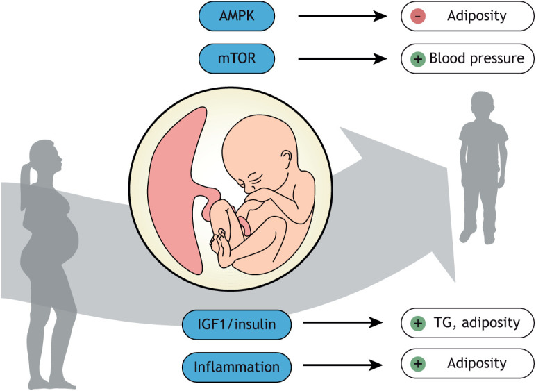
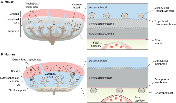
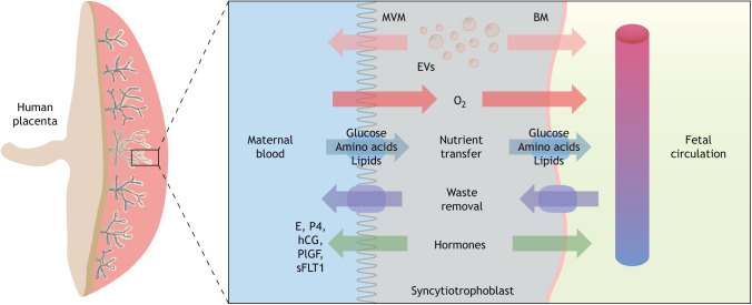
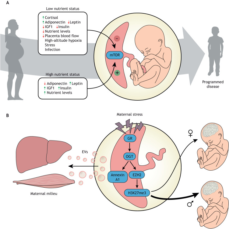
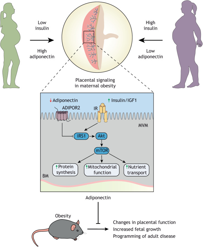
Compelling epidemiological and animal experimental data demonstrate that cardiometabolic and neuropsychiatric diseases originate in a suboptimal intrauterine environment. Here, we review evidence suggesting that altered placental function may, at least in part, mediate the link between the maternal environment and changes in fetal growth and development. Emerging evidence indicates that the placenta controls the development and function of several fetal tissues through nutrient sensing, modulation of trophoblast nutrient transporters and by altering the number and cargo of released extracellular vesicles. In this Review, we discuss the development and functions of the maternal-placental-fetal interface (in humans and mice) and how cross-talk between these compartments may be a mechanism for in utero programming, focusing on mechanistic target of rapamycin (mTOR), adiponectin and O-GlcNac transferase (OGT) signaling. We also discuss how maternal diet and stress influences fetal development and metabolism and how fetal growth restriction can result in susceptibility to developing chronic disease later in life. Finally, we speculate how interventions targeting placental function may offer unprecedented opportunities to prevent cardiometabolic disease in future generations.

Keywords: Epigenetics; Extracellular vesicles; Fetal development; Maternal-fetal exchange; Prenatal; Programming.


Conflict of interest statement

Competing interests The authors declare no competing or financial interests.

Figures

Fig. 1.
Fig. 1.
Key placental metabolic signaling pathways and their associations with cardiometabolic outcomes in children. Placental AMP-activated protein kinase (AMPK) signaling (a cellular energy sensor) is negatively associated with adiposity in children, whereas the activity of placental mechanistic target of rapamycin (mTOR) signaling (a cellular nutrient sensor) is positively correlated to systolic blood pressure at 4-6 years of age. Moreover, placental IGF1/insulin signaling is positively associated with triglyceride (TG) levels and adiposity at 4-6 years of age, and placental inflammation predicts adiposity in children.
Fig. 2.
Fig. 2.
Comparison of mouse and human placental structures. (A,B) Both mouse and human placentas are hemochorial (trophoblast bathed in maternal blood) and discoid in shape. (A) The murine placental structure includes the labyrinth and junctional zones. The box depicts the maternal-fetal interface of the labyrinth, demonstrating the haemotrichorial or three trophoblast layers (a mononuclear trophoblast cell layer and syncytiotrophoblast layers I and II). (B) The human placenta has analogous layers: a decidual layer with spiral arteries and the placental villi. The box details a section of a chorionic villus illustrating the haemomonochorial or monolayer of syncytiotrophoblast cells separating maternal and fetal blood. The apical brush border of the human syncytiotrophoblast in contact with maternal blood is referred to as microvillous membrane (MVM) and the opposing plasma membrane juxtaposed to the fetal capillary is referred to as basal membrane (BM). The corresponding apical membrane in the mouse placenta is referred to as the trophoblast plasma membrane (TPM) and is localized to syncytial layer II.
Fig. 3.
Fig. 3.
Human placental morphology and transport across the maternal-fetal interface. Transport across the syncytiotrophoblast occurs via passive and facilitated diffusion, active transport and endo- and exocytosis. Macromolecules and waste products are transported across the syncytiotrophoblast via specialized transporting proteins, channels and exchangers located in the plasma membranes. Extracellular vesicles (EVs) and hormones released by the syncytiotrophoblast cells are released to the maternal and fetal circulations, thereby participating in maternal-fetal cross-talk. BM, basal plasma membrane; E, estrogen; P4, progesterone; hCG, human chorionic gonadotropin; MVM, microvillous membrane; plGF, placental growth factor; sFLT1, soluble fms-like tyrosine kinase.
Fig. 4.
Fig. 4.
Roles of placental mTOR and OGT in maternal and fetal health. (A) Mechanistic target of rapamycin (mTOR) signaling is one of several key placental sensors integrating maternal signals and conveying information on the ability of the maternal supply line to deliver nutrients and oxygen to the placenta. mTOR is a master regulator of placental function including mitochondrial respiration, nutrient transport, protein synthesis and hormone secretion, thereby regulating fetal growth and development and impacting the long-term health of the offspring. Importantly, these regulatory loops can function in response to both maternal overnutrition and undernutrition to regulate fetal growth according to the available resources. (B) Placental OGT serves as a key cellular mechanism that senses available energy levels and dynamically alters placental function via multiple mechanisms to broadly regulate maternal homeostasis and impact transplacental signals important for fetal development. Importantly, OGT controls local trophoblast responses to a changing maternal environment where maternal stress hormones activate the glucocorticoid receptor (GR), reducing OGT levels. OGT is a key regulator of transcriptomic pathways via stabilization of the H3K27 methyl transferase EZH2. Reduced OGT results in decreases in EZH2 and the transcriptional repressive histone mark, H3K27me3. As OGT is X chromosome-linked, this transcriptomic regulation is much tighter in female XX trophoblast cells than male XY cells, resulting in dynamic placental responses and transplacental signals to the male fetus. A separate cellular signaling pathway links OGT to activation of annexin A1, an essential component in extracellular vesicle (EV) loading and secretion. EVs secreted by the placenta into maternal circulation contribute to homeostatic regulation, including maternal glucose levels in pregnancy. OGT, O-linked N-acetylglucosamine (O-GlcNAc) transferase; EZH2, enhancer of zeste homolog 2.
Fig. 5.
Fig. 5.
Maternal adiponectin informs the placenta about maternal nutrition status. High maternal adiponectin in low nutrient status mothers inhibits insulin signaling in the placenta and reduces nutrient transport capacity. The combination of high adiponectin and low insulin limits placental uptake and protects the mother from additional depletion, resulting in a smaller but potentially viable offspring. In the obese mother with low adiponectin and high insulin there is no inhibition of transport, leading to larger placentas, greater transport capacity and the excess supply of nutrients to the fetus, enhancing fetal growth and resulting in large-for-gestational age babies. These studies demonstrate that the placenta is responding to maternal nutrient status and altering delivery of nutrients to the fetus to match maternal stores. Correcting adiponectin in obese mice to normal levels prevents these changes in placental function, fetal overgrowth and developmental programming of cardiometabolic disease, suggesting that targeting the placenta is a therapeutic alternative that should be further investigated.

References

    1. Acosta, O., Ramirez, V. I., Lager, S., Gaccioli, F., Dudley, D. J., Powell, T. L. and Jansson, T. (2015). Increased glucose and placental GLUT-1 in large infants of obese nondiabetic mothers. Am. J. Obstet. Gynecol. 212, e1-e7. 10.1016/j.ajog.2014.08.009 - DOI - PubMed
    1. Adamson, S. L., Lu, Y., Whiteley, K. J., Holmyard, D., Hemberger, M., Pfarrer, C. and Cross, J. C. (2002). Interactions between trophoblast cells and the maternal and fetal circulation in the mouse placenta. Dev. Biol. 250, 358-373. 10.1006/dbio.2002.0773 - DOI - PubMed
    1. Akhaphong, B., Baumann, D. C., Beetch, M., Lockridge, A. D., Jo, S., Wong, A., Zemanovic, T., Mohan, R., Fondevilla, D. L., Sia, M.et al. (2021). Placental mTOR complex 1 regulates fetal programming of obesity and insulin resistance in mice. JCI Insight 6, e149271. 10.1172/jci.insight.149271 - DOI - PMC - PubMed
    1. Aye, I. L., Gao, X., Weintraub, S. T., Jansson, T. and Powell, T. L. (2014). Adiponectin inhibits insulin function in primary trophoblasts by PPARα-mediated ceramide synthesis. Mol. Endocrinol. 28, 512-524. 10.1210/me.2013-1401 - DOI - PMC - PubMed
    1. Aye, I. L., Rosario, F. J., Powell, T. L. and Jansson, T. (2015). Adiponectin supplementation in pregnant mice prevents the adverse effects of maternal obesity on placental function and fetal growth. Proc. Natl. Acad. Sci. USA 112, 12858-12863. 10.1073/pnas.1515484112 - DOI - PMC - PubMed

Publication types