. 2023 Oct 13;9(41):eadg4479.
doi: 10.1126/sciadv.adg4479. Epub 2023 Oct 13.

Molecular basis of ClC-6 function and its impairment in human disease

Affiliations

Molecular basis of ClC-6 function and its impairment in human disease

Bing Zhang et al. Sci Adv. .

Abstract

ClC-6 is a late endosomal voltage-gated chloride-proton exchanger that is predominantly expressed in the nervous system. Mutated forms of ClC-6 are associated with severe neurological disease. However, the mechanistic role of ClC-6 in normal and pathological states remains largely unknown. Here, we present cryo-EM structures of ClC-6 that guided subsequent functional studies. Previously unrecognized ATP binding to cytosolic ClC-6 domains enhanced ion transport activity. Guided by a disease-causing mutation (p.Y553C), we identified an interaction network formed by Y553/F317/T520 as potential hotspot for disease-causing mutations. This was validated by the identification of a patient with a de novo pathogenic variant p.T520A. Extending these findings, we found contacts between intramembrane helices and connecting loops that modulate the voltage dependence of ClC-6 gating and constitute additional candidate regions for disease-associated gain-of-function mutations. Besides providing insights into the structure, function, and regulation of ClC-6, our work correctly predicts hotspots for CLCN6 mutations in neurodegenerative disorders.


Figures

Fig. 1.
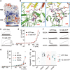
Fig. 1.. Cryo-EM structure of human ClC-6.
(A) Cryo-EM map for the ClC-6 viewed from side (left) and top (right). The two different subunits of the dimer were highlighted by different colors. (B) Cartoon representation of the ClC-6 monomer, colored by structural element. (C) Overall structure of ClC-6 viewed from side (left) and top (right), color codes for subunits were the same as those in (A), and the long disordered region (649 to 677) is circled with red dashed boxes. (D) Structure superimposition of ClC-6 with ClC-7. Color codes ClC-6 and ClC-7 were indicated.
Fig. 2.
Fig. 2.. Electrophysiological characteristics of ClC-6.
(A to C) Whole-cell recording protocol (A) and representative current traces of untransfected Chinese hamster ovary (CHO) cells (B, n = 9/4, indicates independent cells/batches, the same hereinafter) and CHO cells overexpressing WT ClC-6 (C, n = 24/7), with bath solution containing 161 mM Cl (pHo 7.5). After holding at −30 mV, cells were clamped between −100 to +160 mV in 0.8-s steps of 20 mV (A). (D) Current density [current magnitude/cell capacitance (I/C)]-voltage (V) relationship (abbreviated as I/C-V) corresponding to results are shown in (B) and (C). (E to G) Influence of temperature on activation kinetics of ClC-6. Representative current traces at different temperatures (E; 17°, 23°, 29°, 34°C, the corresponding n = 16/4, 12/3, 11/3, and 13/3). The activation time constant (τactivation) at +160 mV of ClC-6 under different temperature was shown as bar graphs (F) and fitted by Arrhenius equation (G). Data were represented as means ± SEM, and one-way ANOVA with post hoc Bonferroni tests (F) was performed. **P < 0.01 and ***P < 0.001.
Fig. 3.
Fig. 3.. Roles of intersubunit and interdomain interactions for ClC-6 function.
(A) Overall structure of ClC-6 with one monomer shown in the surface style. The interaction zones for functional analysis were circled with colored boxes (black: dimeric interactions within TMD; red: N-terminal–mediated polar interactions; blue: C-terminal protruding into opposite CBS domain). (B and C) Magnified view for dimeric interactions within TMD (B) and N-terminal–mediated polar interactions (C). The interaction was shown as dashed line. (D) Representative current traces and τactivation of WT ClC-6 and mutants including E266A (n = 10/3), Q274A (n = 9/3), D52A (n = 9/3), Y53A (n = 9/3), D54A (n = 10/3), N167A (n = 12/3), D240A (n = 12/3), and R828A (n = 12/3). (E) A helix-turn-helix sequence between the CBS domains of one subunit protrudes into the opposite CBS. The cryo-EM density for C terminus was shown as red mesh contoured at 8σ threshold. (F) Magnified view for the C-terminal–mediated interactions. The interactions were shown as dashed lines. (G) Representative current traces and τactivation of ClC-6 mutants including Δ650–674 (n = 10/3), R667A (n = 7/2), and R674A (n = 8/2). Data were represented as means ± SEM, and one-way ANOVA with post hoc Bonferroni tests (D and G) was performed. ***P < 0.001.
Fig. 4.
Fig. 4.. Structure and effect of ATP binding on ClC-6.
(A) Structure of ClC-6 in complex with ATP. ATP-binding site highlighted by yellow dashed box. (B and C) Magnified view of ATP-binding site in ClC-6. The cryo-EM density for ATP molecule shown as blue mesh contoured at 8σ threshold (B). Key residues interacting with ATP shown as sticks (C). (D and E) Representative current traces and I/C-V curves of ClC-6 without or with 10 mM ATP in the pipette solution (n = 9/3). (F and G) Representative current traces and I/C-V curves of mutants predicted to neutralize ATP binding without [empty icons in (G); n = 11/3, 11/3, and 10/3 for R833A, H851A, and H630A] or with 10 mM ATP [filled icons in (G); n = 12/3, 9/3, and 8/2 for R833A, H851A, and H630A) in the pipette solution. (H) Influence of ATP and mutations on the voltage dependence of ClC-6 that was quantified by determining an apparent V1/2. Data were represented as means ± SEM, and unpaired Student’s t tests (E) and two-way ANOVA with post hoc Bonferroni tests (H) were performed. ***P < 0.001 [in (H), compared to WT ClC-6 with an equivalent experimental condition) and #P < 0.05, ##P < 0.01, ###P < 0.001. n.s., not significant.
Fig. 5.
Fig. 5.. Role of Y553/F317/T520 interaction network for the voltage dependence of ClC-6.
(A) Magnified view for the interactions among Y553, F317, and T520. The interactions shown as dashed lines. (B and C) Representative current traces and I/C-V curves of WT ClC-6 and mutants including Y553A (n = 11/3), F317A (n = 12/3). and T520A (n = 7/3). (D) Apparent V1/2 was compared between groups shown in (B) and (C). (E) Value of τactivation at 120, 140, and 160 mV was compared between groups shown in (B) and (C). (F) Representative current traces of WT, Y553W (n = 6/2), and Y553F (n = 11/3). (G) I/C-V curves of WT ClC-6 and mutants including Y553W, Y553F, Y553C, and Y553A. (H) Apparent V1/2 was compared between groups shown in (G). Data were represented as means ± SEM, and one-way ANOVA with post hoc Bonferroni tests (D, E, and H) were performed. ***P < 0.001 (versus WT ClC-6) and ###P < 0.001 (versus Y553C mutant).
Fig. 6.
Fig. 6.. Role of P/Q linker in gating.
(A and B) Representative current traces and I/C-V curves of WT ClC-6, N549A (n = 11/3), and E550A (n = 11/3) mutants. (C) Apparent V1/2 were compared among groups shown in (B). (D) Images displaying GFP fluorescence of CHO cells overexpressing N-terminally GFP-tagged WT ClC-6 and mutants including E550A, Y553A, T520A, and F317A, respectively. Scale bars, 10 μm. (E) Value of τactivation at 120, 140, and 160 mV was compared among Y553A, E550A, and N549A. (F) Magnified view of interactions between N549 and E550. The interactions were shown as dashed lines. Data were represented as means ± SEM, and one-way ANOVA with post hoc Bonferroni tests (C and E) was performed. ***P < 0.001.
Fig. 7.
Fig. 7.. Interactions of the I/J linker with TMD helices modulate ClC-6 activation kinetics.
(A) Surface representation of ClC-6 structure to clarify the position of I/J linker (Yellow ribbon). (B) Magnified view for the interactions between I/J linker and the surrounding residues, the interactions were shown as dashed lines. (C and D) Representative current traces and τactivation of WT ClC-6 and mutants including L311A (n = 9/3), L312A (n = 9/3), F314A (n = 9/3), F454A (n = 8/2) and H455A (n = 9/3). (E) Sequence alignment of the I/J linker and the surrounding residues of ClC-6 with other CLCs. Data represented as means ± SEM; one-way ANOVA with post hoc Bonferroni tests (D) was performed. ***P < 0.001.
Fig. 8.
Fig. 8.. Role of interhelical interactions in ClC-6 gating.
(A) Magnified view for the inter-helical connections between helix O and G, N, and P. Residues that contribute to interhelical contacts were shown as sticks. (B and C) Representative current traces and I/C-V curves of WT ClC-6 and mutants including F489A (n = 9/3), L493A (n = 10/3), and L530A (n = 9/3). (D) Apparent V1/2 were compared between groups shown in (B) and (C). (E and F) Magnified view for the interhelical connections that are far from the Cl binding sites. Residues that contribute to interhelical contacts were shown as sticks. (G and H) Representative current traces and I/C-V curves of mutants including R295A (n = 7/2), Q452A (n = 8/3), R501A (n = 8/3), N505A (n = 8/3), F529A (n = 9/3), and Q446A (n = 5/2). (I) Apparent V1/2 was compared between groups shown in (G) and (H). Data were represented as means ± SEM, and one-way ANOVA with post hoc Bonferroni tests (D and I) was performed. ***P < 0.001.

References

    1. T. J. Jentsch, M. Pusch, CLC chloride channels and transporters: Structure, function, physiology, and disease. Physiol. Rev. 98, 1493–1590 (2018). - PubMed
    1. T. J. Jentsch, Discovery of CLC transport proteins: Cloning, structure, function and pathophysiology. J. Physiol. 593, 4091–4109 (2015). - PMC - PubMed
    1. G. Zifarelli, M. Pusch, CLC chloride channels and transporters: A biophysical and physiological perspective. Rev. Physiol. Biochem. Pharmacol. 158, 23–76 (2007). - PubMed
    1. M. M. Polovitskaya, C. Barbini, D. Martinelli, F. L. Harms, F. S. Cole, P. Calligari, G. Bocchinfuso, L. Stella, A. Ciolfi, M. Niceta, T. Rizza, M. Shinawi, K. Sisco, J. Johannsen, J. Denecke, R. Carrozzo, D. J. Wegner, K. Kutsche, M. Tartaglia, T. J. Jentsch, A recurrent gain-of-function mutation in CLCN6, encoding the ClC-6 Cl/H+-exchanger, causes early-onset neurodegeneration. Am. J. Hum. Genet. 107, 1062–1077 (2020). - PMC - PubMed
    1. T. Y. Chen, Structure and function of CLC channels. Annu. Rev. Physiol. 67, 809–839 (2005). - PubMed