. 2023 Oct 19;83(20):3692-3706.e5.
doi: 10.1016/j.molcel.2023.09.024. Epub 2023 Oct 12.

Sen1 architecture: RNA-DNA hybrid resolution, autoregulation, and insights into SETX inactivation in AOA2

Affiliations

Sen1 architecture: RNA-DNA hybrid resolution, autoregulation, and insights into SETX inactivation in AOA2

C Denise Appel et al. Mol Cell. .

Abstract

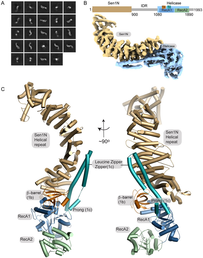
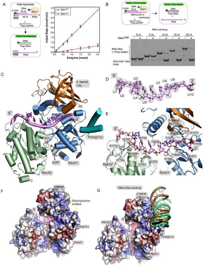
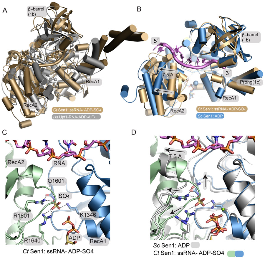
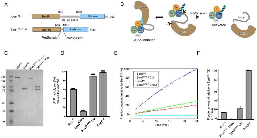
The senataxin (SETX, Sen1 in yeasts) RNA-DNA hybrid resolving helicase regulates multiple nuclear transactions, including DNA replication, transcription, and DNA repair, but the molecular basis for Sen1 activities is ill defined. Here, Sen1 cryoelectron microscopy (cryo-EM) reconstructions reveal an elongated inchworm-like architecture. Sen1 is composed of an amino terminal helical repeat Sen1 N-terminal (Sen1N) regulatory domain that is flexibly linked to its C-terminal SF1B helicase motor core (Sen1Hel) via an intrinsically disordered tether. In an autoinhibited state, the Sen1Sen1N domain regulates substrate engagement by promoting occlusion of the RNA substrate-binding cleft. The X-ray structure of an activated Sen1Hel engaging single-stranded RNA and ADP-SO4 shows that the enzyme encircles RNA and implicates a single-nucleotide power stroke in the Sen1 RNA translocation mechanism. Together, our data unveil dynamic protein-protein and protein-RNA interfaces underpinning helicase regulation and inactivation of human SETX activity by RNA-binding-deficient mutants in ataxia with oculomotor apraxia 2 neurodegenerative disease.

Keywords: DNA repair; Helicase; R-loop; RNA-DNA hybrid; SETX; SF1B; Sen1; X-ray crystallography; cryo-EM; senataxin; transcription.


Conflict of interest statement

Declaration of interests The authors declare no competing interests.

Figures

Figure 1
Figure 1. Cryo-EM structure of Sen1FL
(A) 2D cryo-EM class averages of full-length Sen1 show an asymmetric elongated assembly. (B) Cryo-EM volume of Sen1FL is displayed showing the Sen1N domain in brown, and the helicase core in blue. (C) The Sen1N helical repeat (tan). Extensive interactions with helical repeat are mediated by RecA1 1b (orange) and 1c (turquoise) insertion elements. The LZZ (RecA1 1c insertion) reinforces the interface.
Figure 2
Figure 2. Crystal structure of the Sen1Hel–ssRNA–ADP–SO4 complex
(A) Left: RNA-DNA unwinding substrates. Right: Sen1FL and Sen1Hel at the amounts indicated were incubated with 50 nmol of Sub7 (Table S2). Reactions were initiated by addition of a mixture containing ATP (1 mM) and MgCl2 (2 mM). FAM signal was detected at 520 nm every 10 sec over 30 min at 25 °C. Initial rates were determined by linear fit of the first 5 min of the unwinding reaction. Error bars are SD from 5 replicates. (B) Top: gel-based RNA-DNA unwinding assay. Bottom: Effect of RNA overhang length on Sen1Hel RNA-DNA unwinding activity. Sub1-Sub5 (Supplementary Table 3) at 20 nM, Sen1Hel (50 nM), 19-Trap DNA (Table 1) at 200 nM, ATP (1 mM), MgCl2 (2 mM) were incubated for 15 min at 37 °C. (C) X-ray structure of the Sen1Hel–RNA–ADP–SO4 complex. The extended LZZ coiled coil is not shown. (D) Composite Omit 2Fo-Fc electron density at 1.0 σ, carved 2.0 Å around the RNA chain. (E) Sen1Hel RNA-protein interactions show an extensive RNA binding interface. (F) APBS surface electrostatic surface representation (blue=electropositive, red= electronegative) of the Sen1 RNA binding surface. Sen1 encircles the RNA via its RecA1 and RecA2 domains. (G) Representative HADDOCK RNA-DNA poses displayed show secondary putative RNA-DNA hybrid binding electropositive surface flanks the ssRNA binding site and is assembled by the 1b and 1c RecA1 insertions.
Figure 3.
Figure 3.. A phosphate sensor is a lynchpin in the Sen1 power stroke
(A) Structural superposition of the Sen1Hel–RNA–ADP–SO4 complex (tan) with the HsUpf1-RNA-ADP-AlF4 complex (grey). (B) Structural superposition of the Sen1-RNA-ADP-S04 complex with ScSen1-ADP-Mg2+ complex, RSCB:5MZN. Close superposition of the RecA1 domains is observed. A 7.5 Å translocation is coincident with phosphate release. The blue sphere marks the position of Mg2+ in the ScSen1 -ADP-Mg2+ complex. (C) Sen1 active site. Four residues from the RecA1 and RecA2 domains bind SO4, a mimic of the hydrolyzed phosphate prior to phosphate release. (D) Active site structural superposition of ScSen1-ADP and CtSen1-RNA-ADP-SO4 complexes colored as indicated. Arrows show structural rearrangements of conserved gamma-phosphate sensing residues upon phosphate (SO4 mimic) release.
Figure 4.
Figure 4.. The Sen1N domain is autoinhibitory.
(A) Design of a PreScission Protease cleavable Sen1. (B) Schematic of activation of PreScission Protease cleavable Sen1N-PP-C. (C) Purified Sen1 proteins or PreScission Protease cleaved protein (Sen1N-PP-C – Cut) used in panels D–F. (D) Substrate stimulated ATP hydrolysis of mutant Sen1 proteins was monitored in a phosphate release assay. Sen1FL, Sen1N-PP-C (Uncut), PreScission Protease-cleaved Sen1N-PP-C (Cut) and Sen1Hel (5 nM) were incubated with Sub6 (Supplementary Table 3) at 5 μM and ATP (1 mM) and MgCl2 (2 mM) for 15 min at 37 °C. Error bars are SD from 3 replicates, ***p<0.001, **** p<0.0001. (E) Helicase assay of indicated Sen1 protein (1 nM) was performed as in Figure 2A and normalized to unwound fraction of Sen1Hel for comparison. (F) Comparison of unwound fractions at 25 min from experiments in “E”. Error bars are SD from 3 replicates. Statistical analysis was done using paired t-test relative to Sen1FL. *p<0.05, ***p<0.001, ****p<0.0001.
Figure 5.
Figure 5.. Cryo-EM structure of Sen1N-PP-C
(A) Local resolution of Sen1N-PP-C, calculated in using the Local Resolution Estimation in CryoSparc, and displayed using a blue (high-resolution, 3.17 Å) to red (low-resolution, 4.0 Å) color-coded surface. (B) Three interaction interfaces (Int1-Int3) mediate Sen1Sen1N interaction with Sen1Hel. The LZZ from Sen1Hel binds both Int1 and Int3 on Sen1Sen1N. A ΔLZZ internal deletion construct is marked. (C) Consurf analysis of the Sen1Sen1N surface reveals conserved surface interaction interfaces (Int1-Int3, dotted lines) are involved in interactions with Sen1Hel. Top: Sen1N domain interaction interfaces that bind the helicase domain. Bottom: Helicase domain interaction surfaces that bind the Sen1N domain. (D) Molecular details of the Int1 interdomain Sen1Sen1N - Sen1Hel interaction interface. (E) Cryo-EM density for Sen1Sen1N Int2 region displayed contoured at 11.0 σ overlaid upon a molecular surface diagram of the β-barrel domain (orange). (F) Molecular details of the Int3 interdomain Sen1Sen1N - Sen1Hel interaction interface. (G) MBP-pulldowns show the LZZ is critical for the Sen1Sen1N–Sen1Hel interaction. (H) Trans inhibition of Sen1Hel by Sen1Sen1N. Sen1Hel (1 nM) was pre-incubated with Sen1Sen1N at the indicated concentrations for 5 min on ice. Reactions were initiated by addition of 50 nM Sub7 (Supplementary Table 3) and a mixture containing ATP (1 mM) and MgCl2 (2 mM). The percentage of unwound ssDNA was calculated from the FAM signal at 30 min. Error bars are SD from 3 replicates, **p<0.01, *p<0.05.
Figure 6.
Figure 6.. Mechanism of Sen1 auto-inhibition
(A) Structural superposition of Sen1N-PP-C and the Sen1Hel RNA complex reveals a cascade of conformational rearrangements associated with autoinhibition. (B) Surface diagram of the Sen1-RNA complex and Sen1N-PP-C shows alterations in the RNA binding channel in the closed state “C”. (D) Fluorescence polarization ssRNA binding. Binding to ssRNA was conducted using enzyme titration and monitoring fluorescence polarization from Sub8 (Supplementary Table 3. Error bars reflect SD from 3 replicates. (E) Superposition of an example HADDOCK docking binding pose and the autoinhibited Sen1N-PP-C state.
Figure 7
Figure 7. AOA2 mutants Sen1 RNA binding mechanism
(A) Structural overlay of an Alphafold model of human SETX helicase domain (blue) with the CtSen1 RNA complex (tan). (B) Alphafold model of the hSETX helicase domain showing the location of mapped AOA2 and ALS4 mutations (C) Two AOA2 mutations map to the RNA binding cleft. Human equivalent positions are shown below the CtSen1 numbering in parentheses and the CtSen1 structure is displayed. (D) Fluorescence polarization ssRNA binding. Binding to ssRNA was conducted using enzyme titration and monitoring fluorescence polarization from Sub8 (Supplementary Table 3). Error bars reflect SD from 3 replicates. (E) ATPase activity. Proteins (5 nM) were incubated with Sub6 (Supplementary Table 3) and ATP (1 mM) at 5 μM for 15 min at 37 °C. The L1551W mutant had no measurable activity. Error bars are SD from 3 replicates, **p<0.01, ****p<0.0001. (F) RNA—DNA unwinding activity. Proteins (1 nM) were incubated with 50 nM Sub7 (Supplementary Table 3), ATP (1 mM) and MgCl2 (2 mM). FAM signal at 10 min was used to compare the unwinding activities of Sen1Hel mutants. The L1551W mutant had no measurable activity. Error bars are SD from 3 replicates, **p<0.01, ****p<0.0001. (G) Purified Sen1Hel proteins used in panels B–D. (H) Model for Sen1 autoinhibition and RNA translocation.

References

    1. Garcia-Muse T, and Aguilera A (2019). R Loops: From Physiological to Pathological Roles. Cell 179, 604–618. 10.1016/j.cell.2019.08.055. - DOI - PubMed
    1. Crossley MP, Bocek M, and Cimprich KA (2019). R-Loops as Cellular Regulators and Genomic Threats. Mol Cell 73, 398–411. 10.1016/j.molcel.2019.01.024. - DOI - PMC - PubMed
    1. Groh M, Albulescu LO, Cristini A, and Gromak N (2017). Senataxin: Genome Guardian at the Interface of Transcription and Neurodegeneration. J Mol Biol 429, 3181–3195. 10.1016/j.jmb.2016.10.021. - DOI - PubMed
    1. Ursic D, Chinchilla K, Finkel JS, and Culbertson MR (2004). Multiple protein/protein and protein/RNA interactions suggest roles for yeast DNA/RNA helicase Sen1p in transcription, transcription-coupled DNA repair and RNA processing. Nucleic Acids Res 32, 2441–2452. - PMC - PubMed
    1. Steinmetz EJ, Warren CL, Kuehner JN, Panbehi B, Ansari AZ, and Brow DA (2006). Genome-wide distribution of yeast RNA polymerase II and its control by Sen1 helicase. Mol Cell 24, 735–746. - PubMed

Publication types