Review
. 2024 Jan 24:19:345-370.
doi: 10.1146/annurev-pathmechdis-051222-120750. Epub 2023 Oct 13.

Neurodegenerative Disease Tauopathies

Affiliations
Review

Neurodegenerative Disease Tauopathies

Benjamin C Creekmore et al. Annu Rev Pathol. .

Abstract

Tauopathies are a diverse group of progressive and fatal neurodegenerative diseases characterized by aberrant tau inclusions in the central nervous system. Tau protein forms pathologic fibrillar aggregates that are typically closely associated with neuronal cell death, leading to varied clinical phenotypes including dementia, movement disorders, and motor neuron disease. In this review, we describe the clinicopathologic features of tauopathies and highlight recent advances in understanding the mechanisms that lead to spread of pathologic aggregates through interconnected neuronal pathways. The cell-to-cell propagation of tauopathy is then linked to posttranslational modifications, tau fibril structural variants, and the breakdown of cellular protein quality control.

Keywords: AD; Alzheimer's disease; FTLD; MAPT; cryo-EM; cryogenic electron microscopy; frontotemporal lobar degeneration; microtubule-associated protein tau; proteostasis; tauopathy.


Conflict of interest statement

DISCLOSURE STATEMENT

E.B.L. and B.C.C. are listed as coinventors on a provisional patent titled “Small Molecule VCP Activators and Methods of Use.”

Figures

Figure 1
Figure 1
Pathogenic variants and major splicing variants of tau. Tau is encoded by the MAPT gene on chromosome 17 in humans that contains 16 exons of which exons 1, 4, 5, 7, 9, 11, 12, and 13 are constitutively included. There are six major isoforms in the human brain produced via alternative splicing (7). Splicing of exons 2 and 3 regulates the inclusion of 0, 1, or 2 N-terminal inserts (N1: orange, N2: yellow) into the N-terminal region (red). Alternative splicing of exon 10 (dark pink) creates isoforms with either three or four microtubule binding region (MTBR) repeats (R1: light pink, R2: dark pink, R3: salmon, R4: purple). Tau also has a proline-rich region (blue) and a C-terminal region (white). Some mutations in MAPT lead to pathogenic amino acid changes or alterations in the ratio of tau isoforms (133). Pathogenic intronic variants in MAPT are primarily found in intron 10 and affect the isoform ratio. Pathogenic exonic variants primarily cluster in the MTBR that is responsible for microtubule binding and makes up the tau fibril core.
Figure 2
Figure 2
Posttranslational modifications (PTMs) of disease-relevant tau fibrils. Sarkosyl-insoluble tau fibrils are found in cognitively normal individuals with primary age-related tauopathy (PART) and cognitively impaired individuals with Alzheimer’s disease (AD) or corticobasal degeneration (CBD). The core of these fibrils is made of part of the microtubule binding region (pink/purple) and C-terminal region (white). These different fibrils are variably modified by PTMs including acetylation (purple), ubiquitination (green), phosphorylation (dark gray), and methylation (light blue) (20, 22).
Figure 3
Figure 3
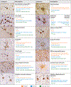
Immunohistochemistry of various tau inclusions, including examples of a neurofibrillary tangle (NFT), Pick body, tufted astrocyte, pretangle, coiled body, thorn-shaped astrocyte, globular astrocytic inclusion, neuritic plaque, ballooned neuron, ramified astrocyte, globose-shaped NFT, astrocytic plaque, argyrophilic grain, granular/fuzzy astrocyte, and globular oligodendrocytic inclusion, found in various tauopathies.
Figure 4
Figure 4
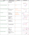
Disease-associated tau fibril cryogenic electron microscopy structures. Diseases with a high-resolution fibril structure are listed with pathologic inclusions found and tau isoform present by immunohistochemistry. Fibril structures are shown for Alzheimer’s disease [Protein Data Bank (PDB) number: 5O3L], primary age-related tauopathy (PDB: 5O3L), chronic traumatic encephalopathy [PDB: 6NWP, Electron Microscopy Data Bank (EMDB) number: 0527], Pick’s disease (PDB: 6GX5), corticobasal degeneration (PDB: 6TJX, EMDB: 10514), argyrophilic grain disease (PDB: 7P6D, EMDB: 13226), aging-related tau astrogliopathy (PDB: 7P6D, EMDB: 13226), progressive supranuclear palsy (PDB: 7P65, EMDB: 13218), and globular glial tauopathy (PDB: 7P67, EMDB: 13220) with the core region colored by tau domain (R1: light pink, R2: dark pink, R3: salmon, R4: purple, C-terminal region: gray) and other notable internal cavity densities highlighted (black). Images created using UCSF ChimeraX, developed by the Resource for Biocomputing, Visualization, and Informatics at the University of California, San Francisco, with support from National Institutes of Health R01-GM129325 and the Office of Cyber Infrastructure and Computational Biology, National Institute of Allergy and Infectious Diseases (79).
Figure 5
Figure 5
Schematic of tau aggregate clearance pathways. For the ubiquitin-proteasome system (UPS) and macroautophagy, ubiquitin is added to aggregates via a cascade of ubiquitin-activating enzymes (E1s) and ubiquitin-conjugating enzymes (E2s), culminating in a transfer of ubiquitin to an aggregate mediated by an E3 ubiquitin ligase. (Left) For the UPS pathway, components of the aggregate are either directly degraded by the proteasome or a protein such as valosin-containing protein (VCP) unfolds and removes a component of the aggregate that is then degraded via the proteasome. (Center) For macroautophagy, p62 delivers ubiquitinated aggregates to a phagophore that becomes an autophagosome and ultimately merges with a lysosome for aggregate degradation. (Right) Chaperone-mediated autophagy is ubiquitin independent and involves the heat shock cognate 70 (Hsc70) complex binding to a KFERQ-like motif. The Hsc70 complex is then targeted to the receptor lysosome-associated membrane protein type 2A (LAMP2A) on a lysosome for internalization and degradation of the aggregate. Figure adapted from Reference .

References

    1. Kovacs GG, Ghetti B, Goedert M. 2022. Classification of diseases with accumulation of Tau protein. Neuropathol. Appl. Neurobiol 48(3):e12792. - PMC - PubMed
    1. Braak H, Alafuzoff I, Arzberger T, Kretzschmar H, Del Tredici K. 2006. Staging of Alzheimer disease-associated neurofibrillary pathology using paraffin sections and immunocytochemistry. Acta Neuropathol. 112(4):389–404 - PMC - PubMed
    1. Braak H, Thal DR, Ghebremedhin E, Del Tredici K. 2011. Stages of the pathologic process in Alzheimer disease: age categories from 1 to 100 years. J. Neuropathol. Exp. Neurol 70(11):960–69 - PubMed
    1. Cho H, Choi JY, Hwang MS, Kim YJ, Lee HM, et al. 2016. In vivo cortical spreading pattern of tau and amyloid in the Alzheimer disease spectrum. Ann. Neurol 80(2):247–58 - PubMed
    1. Schöll M, Lockhart SN, Schonhaut DR, O’Neil JP, Janabi M, et al. 2016. PET imaging of Tau deposition in the aging human brain. Neuron 89(5):971–82 - PMC - PubMed

LinkOut - more resources