. 2023 Nov 8;43(45):7657-7667.
doi: 10.1523/JNEUROSCI.1153-23.2023. Epub 2023 Oct 13.

Impact and Role of Hypothalamic Corticotropin Releasing Hormone Neurons in Withdrawal from Chronic Alcohol Consumption in Female and Male Mice

Affiliations

Impact and Role of Hypothalamic Corticotropin Releasing Hormone Neurons in Withdrawal from Chronic Alcohol Consumption in Female and Male Mice

Sofia Neira et al. J Neurosci. .

Abstract

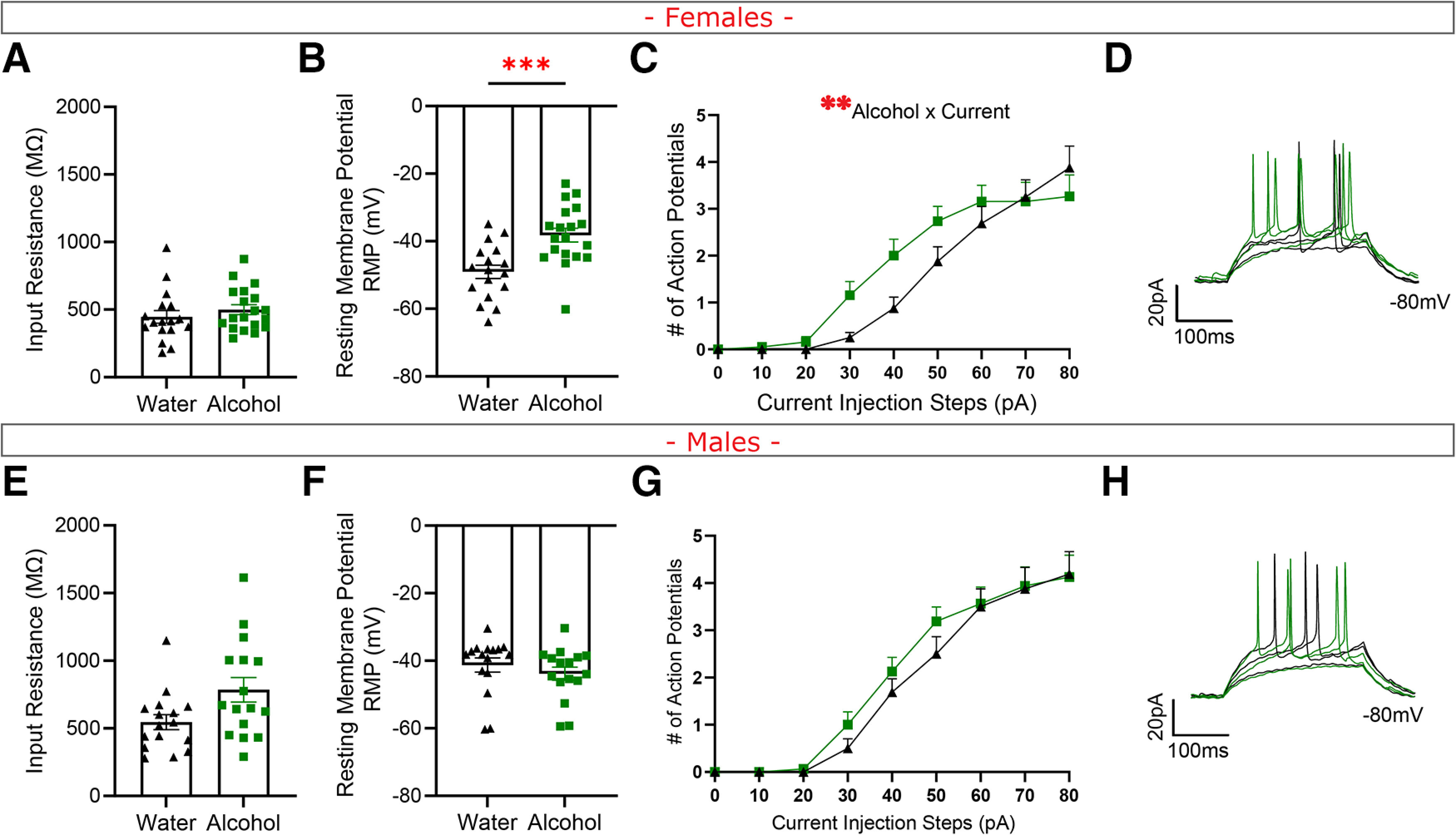
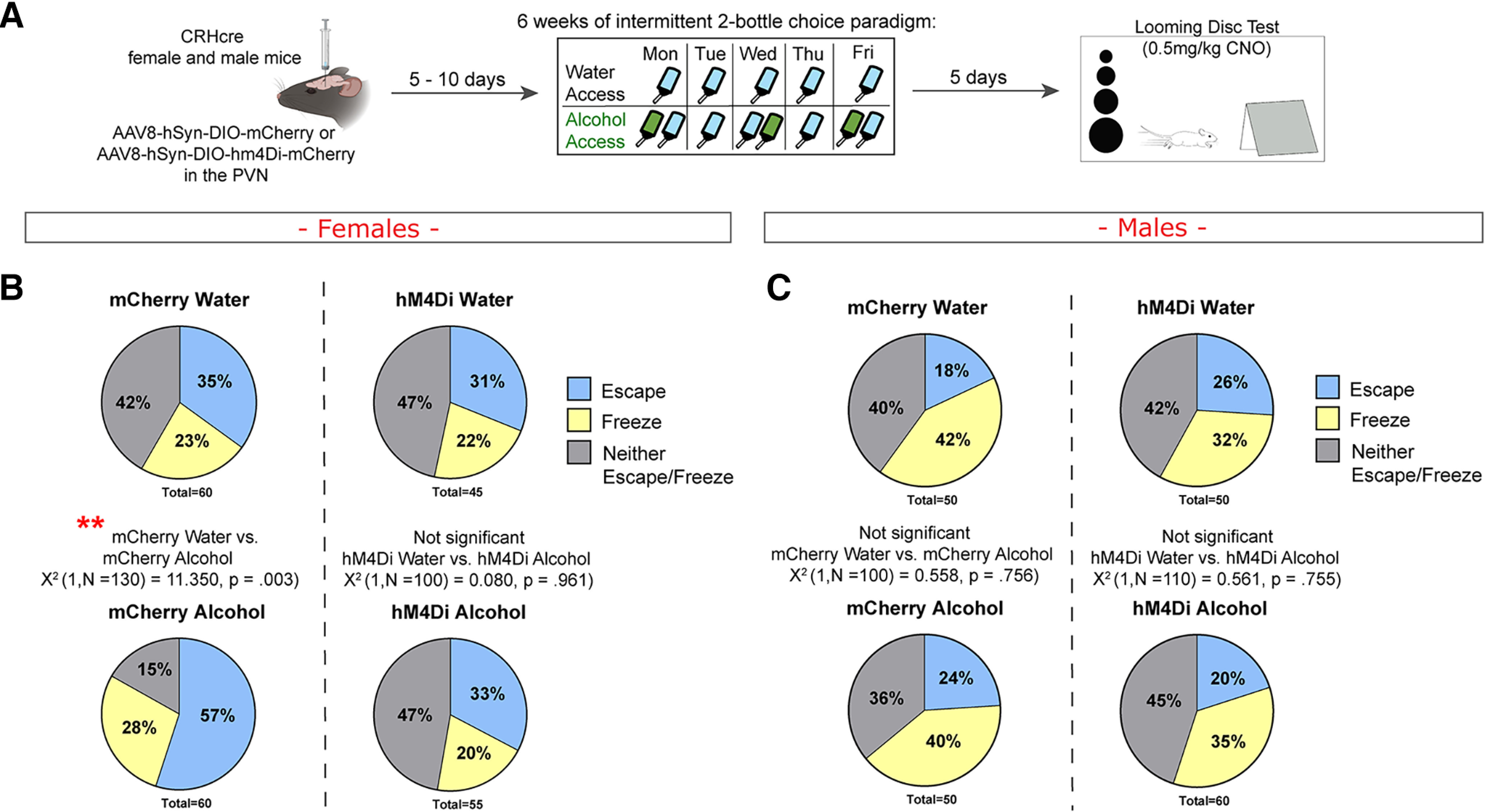
Worldwide, alcohol use and abuse are a leading risk of mortality, causing 5.3% of all deaths (World Health Organization, 2022). The endocrine stress system, initiated by the peripheral release of corticotropin releasing hormone (CRH) from primarily glutamatergic neurons in the paraventricular nucleus of the hypothalamus (PVN), is profoundly linked with alcohol use, abuse, and relapse (Blaine and Sinha, 2017). These PVN CRH-releasing (PVNCRH) neurons are essential for peripheral and central stress responses (Rasiah et al., 2023), but little is known about how alcohol affects these neurons. Here, we show that two-bottle choice alcohol consumption blunts the endocrine-mediated corticosterone response to stress during acute withdrawal in female mice. Conversely, using slice electrophysiology, we demonstrate that acute withdrawal engenders a hyperexcitable phenotype of PVNCRH neurons in females that is accompanied by increased glutamatergic transmission in both male and female mice. GABAergic synaptic transmission was unaffected by alcohol history. We then tested whether chemogenetic inhibition of PVNCRH neurons would restore stress response in female mice with a history of alcohol drinking in the looming disk test, which mimics an approaching predator threat. Accordingly, inhibition of PVNCRH neurons reduced active escape in hM4Di alcohol history mice only. This study indicates that stress-responsive PVNCRH neurons in females are particularly affected by a history of alcohol consumption. Interestingly, women have indicated an increase in heavy alcohol use to cope with stress (Rodriguez et al., 2020), perhaps pointing to a potential underlying mechanism in alcohol-mediated changes to PVNCRH neurons that alter stress response.SIGNIFICANCE STATEMENT Paraventricular nucleus of the hypothalamus neurons that release corticotropin releasing hormone (PVNCRH) are vital for stress response. These neurons have been understudied in relation to alcohol and withdrawal despite profound relations between stress, alcohol use disorders (AUD), and relapse. In this study, we use a variety of techniques to show that acute withdrawal from a history of alcohol impacts peripheral stress response, PVNCRH neurons, and behavior. Specifically, PVNCRH are in a hyperactive state during withdrawal, which drives an increase in active stress coping behaviors in female mice only. Understanding how alcohol use and withdrawal affects stress responding PVNCRH neurons may contribute to finding new potential targets for the treatment of alcohol use disorder.

Keywords: alcohol; behavior; electrophysiology; hypothalamus; mice; withdrawal.


Figures

Figure 1.
Figure 1.
Acute stress in alcohol withdrawal blunts plasma corticosterone and PVNCRH Fos mRNA expression in female alcohol history mice. In females: A, Experimental design for basal measurements after 2-BC alcohol. B, Corticosterone. C, Total number of Fos-positive cells. D, Total number of Crh-positive cells. E, Percent of Crh-positive cells that were also positive for Fos. F, Representative images of water (left) and alcohol (right) mice. G, Experimental design for acute stress measurements after 2-BC alcohol. H, Corticosterone. I, Total number of Fos-positive cells. J, Total number of Crh-positive cells. K, Percent of Crh-positive cells that were also positive for Fos. L, Representative images of water (left) and alcohol (right) mice. In males: M, Experimental design for basal measurements after 2-BC alcohol. N, Corticosterone. O, Total number of Fos-positive cells. P, Total number of Crh-positive cells. Q, Percent of Crh-positive cells that were also positive for Fos. R, Representative images of water (left) and alcohol (right) mice. S, Experimental design for acute stress measurements after 2-BC alcohol. T, Corticosterone. U, Total number of Fos-positive cells. V, Total number of Crh-positive cells. W, Percent of Crh-positive cells that were also positive for Fos. X, Representative images of water (left) and alcohol (right) mice. Data are mean ± SE. B,C,D,H,I,J,N,O,P,T,U,V student's unpaired t-test. E,K,Q,W Mann-Whitney two-tailed test. * p < 0.05, ** p < 0.01.
Figure 2.
Figure 2.
Chronic alcohol consumption engenders a hyperexcitable phenotype in PVNCRH neurons in female alcohol withdrawal mice. In females: A, Input resistance. B, RMP in mV. C, Current step injection plot. D, Representative 30-50 pA current steps in current step injection plot. In males: E, Input resistance. F, RMP in mV. G, Current step injection plot. H, Representative 30-50 pA current steps in current step injection plot. Data are mean ± SE. A, B, E, F, nested t-test. C, G, Two-way repeated-measures ANOVA. **p < 0.01. ***p < 0.001.
Figure 3.
Figure 3.
A history of alcohol increases glutamatergic synaptic transmission in female and male alcohol withdrawal mice. In females: A, mEPSC frequency. B, mEPSC amplitude. C, Representative traces for water (black) and alcohol (green) mEPSCs. D, sEPSC frequency. E, sEPSC amplitude. F, sEPSC amplitude. Representative traces for water (black) and alcohol (green) sEPSCs. G, mIPSC frequency. H, mIPSC amplitude. I, Representative traces for water (black) and alcohol (green) mIPSCs. J, sIPSC frequency. K, sIPSC amplitude. L, Representative traces for water (black) and alcohol (green) sIPSCs. In males: M, mEPSC frequency. N, mEPSC amplitude. O, Representative traces for water (black) and alcohol (green) mEPSCs. P, sEPSC frequency. Q, sEPSC amplitude. R, Representative traces for water (black) and alcohol (green) sEPSCs. S, mIPSC frequency. Data are mean ± SE. T, mIPSC amplitude. U, Representative traces for water (black) and alcohol (green) mIPSCs. V, sIPSC frequency. W, sIPSC amplitude. X, Representative traces for water (black) and alcohol (green) sIPSCs. *p < 0.05; **p < 0.01; nested t-test.
Figure 4.
Figure 4.
hM4Di-mediated chemogenetic inhibition of PVNCRH neurons did not affect mouse responses to the home-cage or free social interaction test in acute withdrawal from chronic alcohol. A, Experimental design for female and male CRH-cre mice. B, Virus placements. Each color represents 1 mouse. In females: C, Fifteen minute home-cage test. D, Distance moved. E, Rear time. F, Dig time. G, Groom time. H, Ten minute free social interaction test. I, Face contact. J, Anogenital contact. K, Allogrooming. L, Fighting. In males: M, Fifteen minute home-cage test. N, Distance moved. O, Rear time. P, Dig time. Q, Groom time. R, Ten minute free social interaction test. S, Face contact. T, Anogenital contact. U, Allogrooming. V, Fighting. Data are mean ± SE. Two-way repeated measures ANOVA. #Main effect of hM4Di virus. $Main effect of alcohol history.
Figure 5.
Figure 5.
hM4Di-mediated chemogenetic inhibition of PVNCRH neurons reduces looming disk active escape responses in female alcohol history mice during withdrawal. A, Experimental design. B, Female escape (blue), freeze (yellow), or neither escape nor freeze (gray) response to loom presentation. C, Male escape (blue), freeze (yellow), or neither escape nor freeze (gray) response to loom presentation. Data are percent escape, freeze, or neither escape/freeze of total looming disc trials. **p < 0.01 (χ2).
Figure 6.
Figure 6.
Working model of PVNCRH neuron alterations in female and male mice. Summary of findings in females (left) and males (right) in withdrawal from a chronic history of alcohol consumption.

References

    1. Adinoff B, Krebaum SR, Chandler PA, Ye W, Brown MB, Williams MJ (2005) Dissection of hypothalamic-pituitary-adrenal axis pathology in 1-month-abstinent alcohol-dependent men: 1. Adrenocortical and pituitary glucocorticoid responsiveness. Alcohol Clin Exp Res 29:517–527. 10.1097/01.alc.0000158940.05529.0a - DOI - PMC - PubMed
    1. Armario A, Vallès A, Dal-Zotto S, Márquez C, Belda X (2004) A single exposure to severe stressors causes long-term desensitisation of the physiological response to the homotypic stressor. Stress 7:157–172. 10.1080/10253890400010721 - DOI - PubMed
    1. Bains JS, Cusulin JIW, Inoue W (2015) Stress-related synaptic plasticity in the hypothalamus. Nat Rev Neurosci 16:377–388. 10.1038/nrn3881 - DOI - PubMed
    1. Bangasser DA, Eck SR, Telenson AM, Salvatore M (2018) Sex differences in stress regulation of arousal and cognition. Physiol Behav 187:42–50. 10.1016/j.physbeh.2017.09.025 - DOI - PMC - PubMed
    1. Bankhead P, Loughrey MB, Fernández JA, Dombrowski Y, McArt DG, Dunne PD, McQuaid S, Gray RT, Murray LJ, Coleman HG, James JA, Salto-Tellez M, Hamilton PW (2017) QuPath: Open Source software for digital pathology image analysis. Sci Rep 7:1. 10.1038/s41598-017-17204-5 - DOI - PMC - PubMed

Publication types

Substances

LinkOut - more resources