. 2023 Oct 13;9(1):374.
doi: 10.1038/s41420-023-01674-w.

MEK inhibitors increase the mortality rate in mice with LPS-induced inflammation through IL-12-NO signaling

Affiliations

MEK inhibitors increase the mortality rate in mice with LPS-induced inflammation through IL-12-NO signaling

Ryota Hashimoto et al. Cell Death Discov. .

Abstract

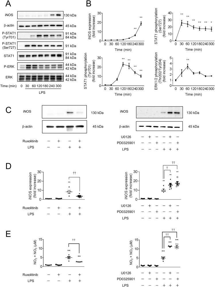
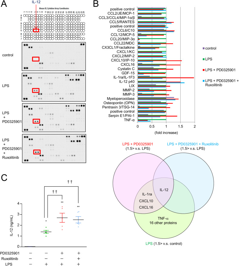
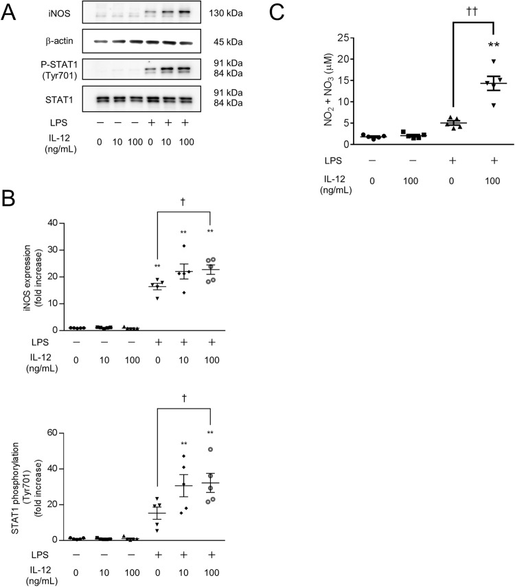
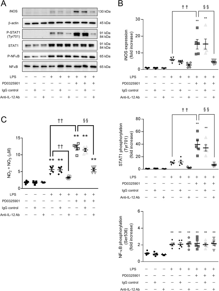
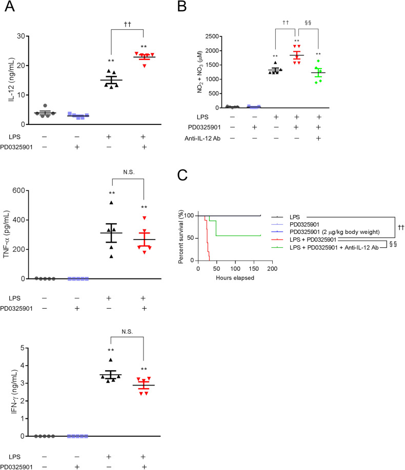
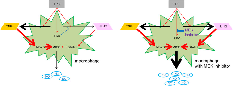
Lipopolysaccharide (LPS) is an endotoxin that can cause an acute inflammatory response. Nitric oxide (NO) is one of the most important innate immune system components and is synthesized by inducible NOS (iNOS) in macrophages in response to stimulation with LPS. LPS activates the RAS-RAF-mitogen-activated protein kinase/ERK kinase (MEK)-extracellular-signal-regulated kinase (ERK) signaling cascade in macrophages. The purpose of this study was to examine how the combination of LPS and MEK inhibitors, which have been used as anticancer agents in recent years, affects inflammation. We showed that MEK inhibitors enhanced iNOS expression and NO production in LPS-stimulated mouse bone marrow-derived macrophages. A MEK inhibitor increased the mortality rate in mice with LPS-induced inflammation. The expression of the cytokine interleukin-12 (IL-12) in macrophages was enhanced by the MEK inhibitor, as shown by a cytokine array and ELISA. IL-12 enhanced iNOS expression and NO production in response to LPS. We also showed that tumor necrosis factor (TNF-α) was secreted by macrophage after stimulation with LPS and that TNF-α and IL-12 synergistically induced iNOS expression and NO production. An anti-IL-12 neutralizing antibody prevented NO production and mortality in an LPS-induced inflammation mouse model in the presence of a MEK inhibitor. These results suggest that the MEK inhibitor increases the mortality rate in mice with LPS-induced inflammation through IL-12-NO signaling.


Conflict of interest statement

The authors declare no competing interests.

Figures

Fig. 1
Fig. 1. LPS-induced iNOS expression in mouse macrophages is suppressed by a JAK-STAT inhibitor and enhanced by a MEK inhibitor.
A, B Bone marrow-derived macrophages (BMDMs) were treated with 100 ng/mL lipopolysaccharide (LPS). The cell extracts were sampled at different time points as indicated after LPS stimulation and analyzed by western blotting. After LPS stimulation, increased phosphorylation of ERK1/2 (Thr202/Tyr204), STAT1 (Ser727), STAT1 (Tyr701), and inducible nitric oxide synthase (iNOS) were detected. n = 5 (*p < 0.05, **p < 0.01 vs. pretreatment (0 min) group). CE BMDMs were treated with inhibitors of JAK-STAT (1 μM ruxolitinib) and MAPK/MEK (1 μM U0126 or 10 nM PD0325901) for 60 min, followed by treatment with 100 ng/mL LPS for 300 min (C, D) or overnight (E). C, D The cell extracts were analyzed by western blotting. LPS-induced iNOS expression was suppressed by a JAK-STAT inhibitor and enhanced by a MEK inhibitor. E The concentration of NO in the supernatant was determined using 2,3-diaminonaphthalene (DAN) and is shown as the concentration of NO2 + NO3. CE n = 5 (**p < 0.01 vs. the control group; p < 0.05, ††p < 0.01 vs. the LPS-treated group).
Fig. 2
Fig. 2. Enhanced iNOS expression and NO production in LPS-stimulated macrophages treated with a MEK inhibitor were blocked by a JAK-STAT inhibitor.
BMDMs were treated with JAK-STAT and MEK inhibitors for 60 min, followed by stimulation with LPS for 60 min, 300 min (AD) or overnight (E). AD The cell extracts were analyzed by western blotting. E The concentration of NO in the supernatant is shown as the concentration of NO2 + NO3. A, B Pretreatment with the MEK inhibitor enhanced the phosphorylation of STAT1 (Tyr701) after stimulation with LPS. CE Enhanced iNOS expression and NO production in response to stimulation with LPS and a MEK inhibitor were blocked by a JAK-STAT inhibitor. AE n = 5 (*p < 0.05, **p < 0.01 vs. the control group; ††p < 0.01 vs. the LPS-treated group; §§p < 0.01).
Fig. 3
Fig. 3. Increased IL-12 secretion in response to stimulation with LPS and a MEK inhibitor.
AC BMDMs were treated with the MEK inhibitor PD0325901 and the JAK-STAT inhibitor ruxolitinib for 60 min, followed by stimulation with LPS for 300 min. A membrane-based multiplex antibody array was used to determine the relative levels of secreted cytokines in the supernatant. A Representative blots of the antibody array are shown. B The bar graph shows the densitometric analysis (n = 1). IL-12 was over 1.5 times more abundant in the LPS + PD0325901 group and the LPS + PD0325901 + ruxolitinib group than in the LPS-treated group. C The concentration of IL-12 in the supernatant was determined by ELISA. n = 5 (*p < 0.05, **p < 0.01 vs. the control group; ††p < 0.01 vs. the LPS-treated group).
Fig. 4
Fig. 4. IL-12 enhances iNOS expression and NO production in LPS-stimulated macrophages.
BMDMs were treated with the indicated concentrations of IL-12 and LPS for 300 min (A, B) or overnight (C). A, B The cell extracts were analyzed by western blotting. C The concentration of NO in the supernatant is shown as the concentration of NO2 + NO3. AC IL-12 enhanced the phosphorylation of STAT1 (Tyr701), iNOS expression, and NO production in response to stimulation with LPS. n = 5 (**p < 0.01 vs. the control group; p < 0.05, ††p < 0.01 vs. the LPS-treated group).
Fig. 5
Fig. 5. IL-12 enhances iNOS expression and NO production in TNF-α−treated macrophages.
A BMDMs were treated with a MEK inhibitor and a JAK-STAT inhibitor for 60 min, followed by stimulation with LPS for 300 min. The concentrations of TNF-α and IFN-γ in the supernatant were determined by ELISA. TNF-α secretion was increased by LPS. BD BMDMs were treated with 100 ng/mL IL-12 in the presence of TNF-α (30 ng/mL), IFN-γ (30 ng/mL), or TNF-α plus IFN-γ for 300 min (B, C) or overnight (D). B, C The cell extracts were analyzed by western blotting. D The concentration of NO in the supernatant is shown as the concentration of NO2 + NO3. BD IL-12 enhanced the phosphorylation of STAT1 (Tyr701), iNOS expression, and NO production in the presence of TNF-α. AD n = 5 (*p < 0.05, **p < 0.01 vs. the control group; p < 0.05, ††p < 0.01). N.S. means not significant, and N.D. means not detected.
Fig. 6
Fig. 6. The increases in iNOS expression and NO production in LPS-stimulated macrophages treated with a MEK inhibitor are blocked by an anti-IL-12 neutralizing antibody.
BMDMs were treated with the MEK inhibitor PD0325901 for 60 min, followed by stimulation with LPS and the indicated concentrations of anti-IL-12 neutralizing antibodies or isotype control antibodies (anti-trinitrophenol antibody) for 300 min (A, B) or overnight (C). A, B The cell extracts were analyzed by western blotting. C The concentration of NO in the supernatant is shown as the concentration of NO2 + NO3. AC The LPS-induced increases in STAT1 phosphorylation (Tyr701), iNOS expression, and NO production were blocked by the anti-IL-12 neutralizing antibody. n = 5 (**p < 0.01 vs. the control group; ††p < 0.01 vs. the LPS-treated group; §§p < 0.01).
Fig. 7
Fig. 7. An anti-IL-12 neutralizing antibody prevents NO production and mortality in an LPS-induced inflammation mouse model in the presence of a MEK inhibitor.
Mice were intraperitoneally injected with PBS (control), LPS (20 mg/kg body weight), the MEK inhibitor PD0325901 (200 ng/kg body weight or 2 μg/kg body weight), LPS plus PD0325901 (200 ng/kg body weight), or LPS, PD0325901 (200 ng/kg body weight), plus an anti-IL-12 neutralizing antibody (38.8 mg/kg body weight). A, B Serum was collected 16 h after intraperitoneal injection. A Serum concentrations of IL-12, TNF-α, and IFN-γ were determined by ELISA. B The concentration of NO in the protein-depleted serum was determined by a method using DAN and is shown as the concentration of NO2 + NO3. A, B n = 5 (**p < 0.01 vs. the control group; ††p < 0.01 vs. the LPS-treated group; §§p < 0.01). N.S. means not significant. C Survival rates of mice after intraperitoneal injection are shown. n = 5 for the PD0325901 (200 ng/kg body weight) and PD0325901 (2 μg/kg body weight) groups, and n = 10 for each other group (††p < 0.01 vs. the LPS-treated group; §§p < 0.01).
Fig. 8
Fig. 8. Schematic overview of macrophage treatment with MEK inhibitors.
Macrophages are the major producers of IL-12 and TNF-α in response to exogenous or endogenous signals, including LPS. In the present study, we revealed that IL-12 increases iNOS expression and NO production in macrophages in the presence of TNF-α in vitro. We suggest that macrophages treated with MEK inhibitors produce abundant NO through IL-12-STAT1 signaling, which results in an increased mortality rate in an LPS-induced inflammation model.

Similar articles

Cited by

References

    1. Kennedy LB, Salama AKS. A review of cancer immunotherapy toxicity. CA Cancer J Clin. 2020;70:86–104. - PubMed
    1. Zhang Y, Zhang Z. The history and advances in cancer immunotherapy: understanding the characteristics of tumor-infiltrating immune cells and their therapeutic implications. Cell Mol Immunol. 2020;17:807–21. - PMC - PubMed
    1. Felgner S, Kocijancic D, Frahm M, Weiss S. Bacteria in cancer therapy: renaissance of an old concept. Int J Microbiol. 2016;2016:8451728. - PMC - PubMed
    1. Mills H, Acquah R, Tang N, Cheung L, Klenk S, Glassen R, et al. The use of bacteria in cancer treatment: a review from the perspective of cellular microbiology. Emerg Med Int. 2022;2022:8127137. - PMC - PubMed
    1. Felgner S, Kocijancic D, Frahm M, Curtiss R, 3rd, Erhardt M, Weiss S. Optimizing Salmonella enterica serovar Typhimurium for bacteria-mediated tumor therapy. Gut Microbes. 2016;7:171–7. - PMC - PubMed