. 2023 Oct 13;13(1):318.
doi: 10.1038/s41398-023-02605-0.

Brain proteomic atlas of alcohol use disorder in adult males

Affiliations

Brain proteomic atlas of alcohol use disorder in adult males

Pang-Ning Teng et al. Transl Psychiatry. .

Abstract

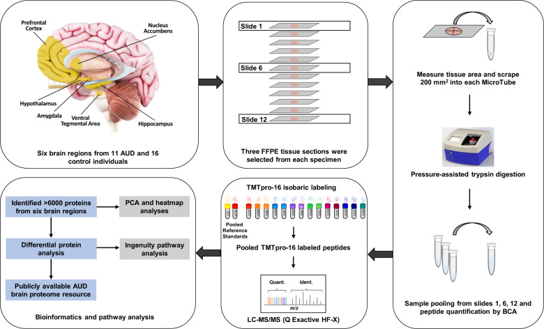
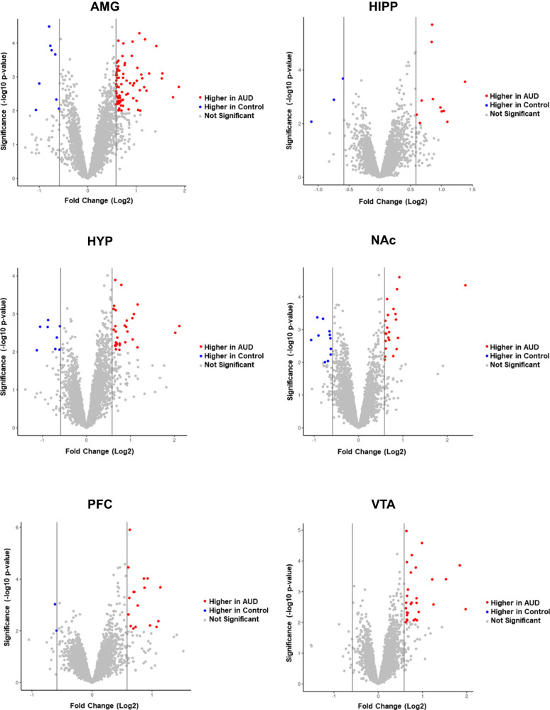
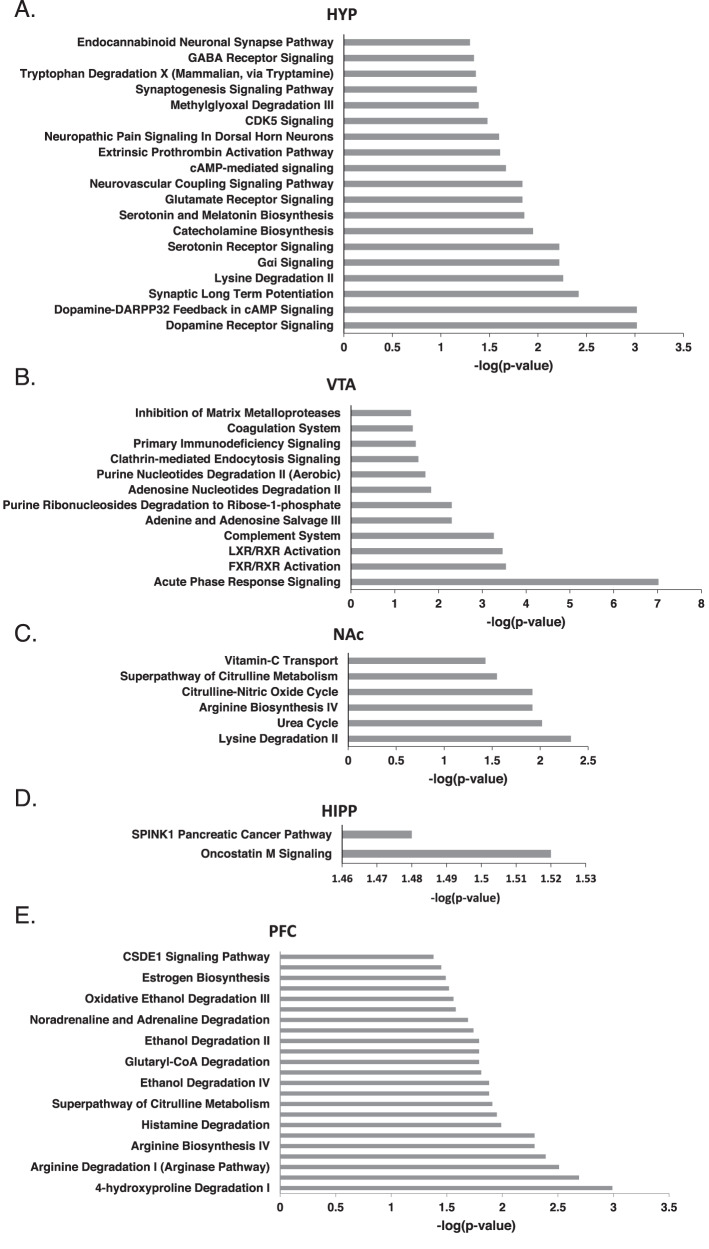
Alcohol use disorder (AUD) affects transcriptomic, epigenetic and proteomic expression in several organs, including the brain. There has not been a comprehensive analysis of altered protein abundance focusing on the multiple brain regions that undergo neuroadaptations occurring in AUD. We performed a quantitative proteomic analysis using a liquid chromatography-tandem mass spectrometry (LC-MS/MS) analysis of human postmortem tissue from brain regions that play key roles in the development and maintenance of AUD, the amygdala (AMG), hippocampus (HIPP), hypothalamus (HYP), nucleus accumbens (NAc), prefrontal cortex (PFC) and ventral tegmental area (VTA). Brain tissues were from adult males with AUD (n = 11) and matched controls (n = 16). Across the two groups, there were >6000 proteins quantified with differential protein abundance in AUD compared to controls in each of the six brain regions. The region with the greatest number of differentially expressed proteins was the AMG, followed by the HYP. Pathways associated with differentially expressed proteins between groups (fold change > 1.5 and LIMMA p < 0.01) were analyzed by Ingenuity Pathway Analysis (IPA). In the AMG, adrenergic, opioid, oxytocin, GABA receptor and cytokine pathways were among the most enriched. In the HYP, dopaminergic signaling pathways were the most enriched. Proteins with differential abundance in AUD highlight potential therapeutic targets such as oxytocin, CSNK1D (PF-670462), GABAB receptor and opioid receptors and may lead to the identification of other potential targets. These results improve our understanding of the molecular alterations of AUD across brain regions that are associated with the development and maintenance of AUD. Proteomic data from this study is publicly available at www.lmdomics.org/AUDBrainProteomeAtlas/ .


Conflict of interest statement

The authors declare no competing interests.

Figures

Fig. 1
Fig. 1. Overview of the experimental workflow.
FFPE tissue from six brain regions of adult AUD (n = 11) and control (n = 16) individuals were harvested for quantitative proteomic analysis.
Fig. 2
Fig. 2. Global brain proteome.
A PCA of global brain proteomics. B Hierarchical clustering of top 100 most variable proteins. C Cellular localization of total proteins identified from each brain region. D Molecular functions of total proteins identified from each brain region. AMG amygdala, HIPP hippocampus, HYP hypothalamus, NAc nucleus accumbens, PFC prefrontal cortex, VTA ventral tegmental area.
Fig. 3
Fig. 3. Volcano plot of proteins with differential abundance between AUD and control.
Differentially expressed proteins (FC cutoff = 1.5, p < 0.01) between AUD and control groups in amygdala (AMG), hippocampus (HIPP), hypothalamus (HYP), nucleus accumbens (NAc), prefrontal cortex (PFC) and ventral tegmental area (VTA).
Fig. 4
Fig. 4. Cellular compartment and molecular functions of differentially expressed proteins between AUD and control groups.
Cellular compartment (A) and molecular function (B) of proteins with differential abundance between AUD and controls (FC cutoff = 1.5, p < 0.01) from all brain regions.
Fig. 5
Fig. 5. Canonical pathway analysis of differentially expressed proteins between AUD and control groups in AMG.
Pathways impacted by AUD in A amygdala (AMG). B activated or inactivated pathways in AMG impacted by AUD. Ingenuity Pathway Analysis showing activated (red bars; positive z-scores) and inactivated pathways (blue bars; negative z-score).
Fig. 6
Fig. 6. Canonical pathway analysis of differentially expressed proteins between AUD and control groups in brain regions.
Pathways impacted by AUD in A hypothalamus (HYP), B ventral tegmental area (VTA), C nucleus accumbens (NAc), D hippocampus (HIPP) and E prefrontal cortex (PFC).

References

    1. Rehm J, Gmel GE, Sr, Gmel G, Hasan OSM, Imtiaz S, Popova S, et al. The relationship between different dimensions of alcohol use and the burden of disease—an update. Addiction. 2017;112:968–1001. doi: 10.1111/add.13757. - DOI - PMC - PubMed
    1. Koob GF, Volkow ND. Neurobiology of addiction: a neurocircuitry analysis. Lancet Psychiatry. 2016;3:760–73. doi: 10.1016/S2215-0366(16)00104-8. - DOI - PMC - PubMed
    1. Rachdaoui N, Sarkar DK. Pathophysiology of the effects of alcohol abuse on the endocrine system. Alcohol Res. 2017;38:255–76. - PMC - PubMed
    1. Gorini G, Harris RA, Mayfield RD. Proteomic approaches and identification of novel therapeutic targets for alcoholism. Neuropsychopharmacology. 2014;39:104–30. doi: 10.1038/npp.2013.182. - DOI - PMC - PubMed
    1. Warden AS, Mayfield RD. Gene expression profiling in the human alcoholic brain. Neuropharmacology. 2017;122:161–74. doi: 10.1016/j.neuropharm.2017.02.017. - DOI - PMC - PubMed

Publication types