Retest variability and patient reliability indices of quantitative fundus autofluorescence in age-related macular degeneration: a MACUSTAR study report
- PMID: 37833348
- PMCID: PMC10576044
- DOI: 10.1038/s41598-023-43417-y
Retest variability and patient reliability indices of quantitative fundus autofluorescence in age-related macular degeneration: a MACUSTAR study report
Abstract
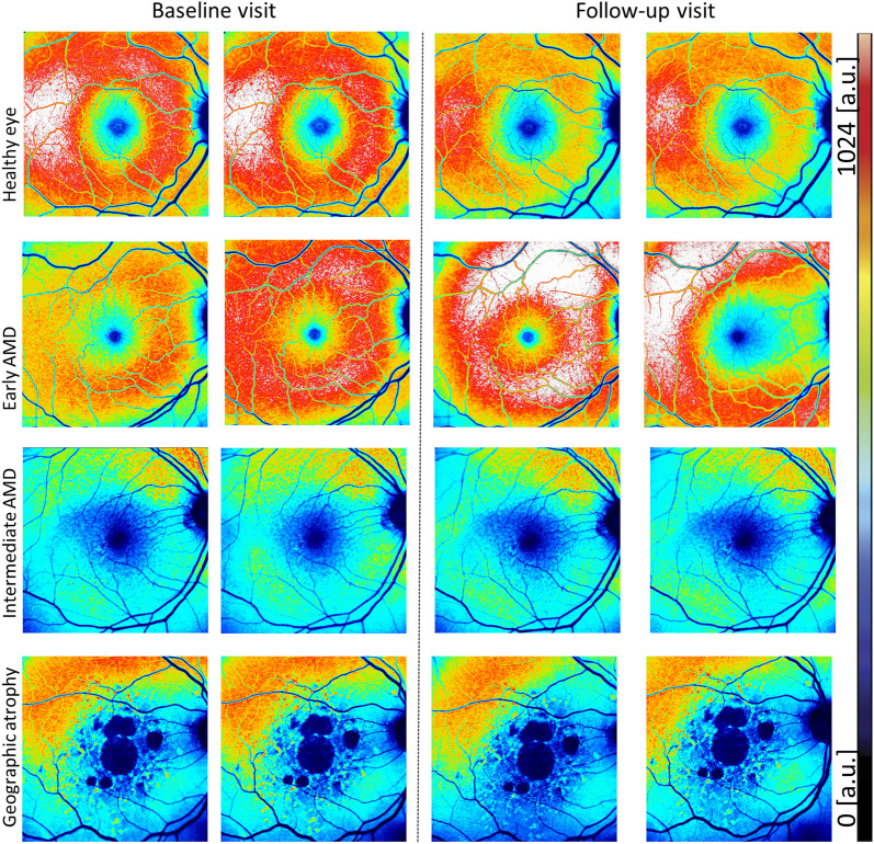
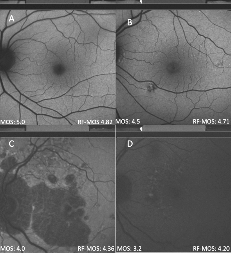
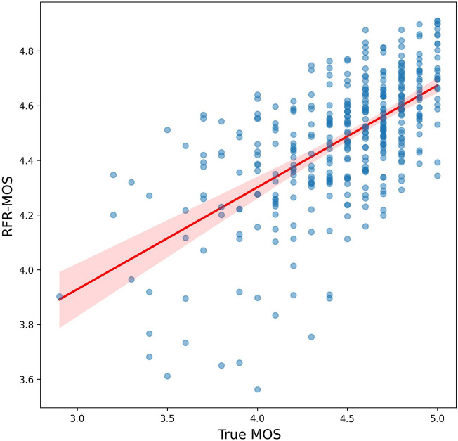
This study aimed to determine the retest variability of quantitative fundus autofluorescence (QAF) in patients with and without age-related macular degeneration (AMD) and evaluate the predictive value of patient reliability indices on retest reliability. A total of 132 eyes from 68 patients were examined, including healthy individuals and those with various stages of AMD. Duplicate QAF imaging was conducted at baseline and 2 weeks later across six study sites. Intraclass correlation (ICC) analysis was used to evaluate the consistency of imaging, and mean opinion scores (MOS) of image quality were generated by two researchers. The contribution of MOS and other factors to retest variation was assessed using mixed-effect linear models. Additionally, a Random Forest Regressor was trained to evaluate the extent to which manual image grading of image quality could be replaced by automated assessment (inferred MOS). The results showed that ICC values were high for all QAF images, with slightly lower values in AMD-affected eyes. The average inter-day ICC was found to be 0.77 for QAF segments within the QAF8 ring and 0.74 for peripheral segments. Image quality was predicted with a mean absolute error of 0.27 on a 5-point scale, and of all evaluated reliability indices, MOS/inferred MOS proved most important. The findings suggest that QAF allows for reliable testing of autofluorescence levels at the posterior pole in patients with AMD in a multicenter, multioperator setting. Patient reliability indices could serve as eligibility criteria for clinical trials, helping identify patients with adequate retest reliability.
© 2023. Springer Nature Limited.
Conflict of interest statement
LvdE: Heidelberg Engineering (R). MM, MV, JH: None. MS: Gerok Research Grant (BONFOR O-137.0030, Faculty of Medicine, University of Bonn, Bonn, Germany), Carl Zeiss MedicTec AG (F), CenterVue (F), Heidelberg Engineering (F). JHT: Heidelberg Engineering (F), Optos (F), Carl Zeiss Meditec (F), CenterVue (F), Novartis (R), Okko (R). KRS: MacRegen Inc (I). SSV: AlphaRET (C), Apellis (C, R), Bayer (F), Bioeq (C), Carl Zeiss MediTec (F), Heidelberg Engineering (F, R), Katairo (C), Kubota Vision (C), Novartis (C, F), Pixium (C), Perceive Therapeutics (C), Roche (C, F), SparingVision (C), STZ GRADE Reading Center (O). RPF: C Alimera, Apellis, Bayer, Böhringer-Ingelheim, Novartis, ODOS, Oxford Innovation, ProGenerika, Roche/Genentech; F Biogen, CentreVue (now Icare), Heidelberg Engineering, Zeiss Meditec. FGH: Acucela (C,F), Allergan (F), Apellis (C, F), Bayer (C, F), Boehringer-Ingelheim (C), Bioeq/Formycon (F,C), CenterVue (F), Ellex (F), Roche/Genentech (C,F), Geuder (C,F), Graybug (C), Gyroscope (C), Heidelberg Engineering (C,F), IvericBio (C, F), Kanghong (C,F), LinBioscience (C), NightStarX (F), Novartis (C,F), Optos (F), Oxurion (C), Pixium Vision (C,F), Oxurion (C), Stealth BioTherapeutics (C), Zeiss (F,C). TA: Roche (C), Novartis (C), Novartis (R), Apellis (C), Bayer (C).
Figures