. 2023 Oct 13;13(1):17361.
doi: 10.1038/s41598-023-44539-z.

The utility of automatic segmentation of kidney MRI in chronic kidney disease using a 3D convolutional neural network

Affiliations

The utility of automatic segmentation of kidney MRI in chronic kidney disease using a 3D convolutional neural network

Kaiji Inoue et al. Sci Rep. .

Abstract

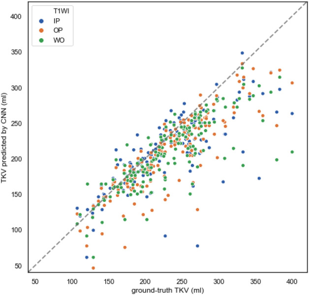
We developed a 3D convolutional neural network (CNN)-based automatic kidney segmentation method for patients with chronic kidney disease (CKD) using MRI Dixon-based T1-weighted in-phase (IP)/opposed-phase (OP)/water-only (WO) images. The dataset comprised 100 participants with renal dysfunction (RD; eGFR < 45 mL/min/1.73 m2) and 70 without (non-RD; eGFR ≥ 45 mL/min/1.73 m2). The model was applied to the right, left, and both kidneys; it was first evaluated on the non-RD group data and subsequently on the combined data of the RD and non-RD groups. For bilateral kidney segmentation of the non-RD group, the best performance was obtained when using IP image, with a Dice score of 0.902 ± 0.034, average surface distance of 1.46 ± 0.75 mm, and a difference of - 27 ± 21 mL between ground-truth and automatically computed volume. Slightly worse results were obtained for the combined data of the RD and non-RD groups and for unilateral kidney segmentation, particularly when segmenting the right kidney from the OP images. Our 3D CNN-assisted automatic segmentation tools can be utilized in future studies on total kidney volume measurements and various image analyses of a large number of patients with CKD.


Conflict of interest statement

The authors declare no competing interests.

Figures

Figure 1
Figure 1
An overview of our three-dimensional convolutional neural network (3D CNN) model used for kidney segmentation. Input is a T1-weighted kidney MRI, followed by 3D convolutional (Conv3D) layers with four convolution blocks in the encoding and decoding branches and a bottleneck convolution block between the two branches. The number of channels is given above each block. The batch normalization (BN) and the parametric rectified linear unit (PReLU) layers are indicated.
Figure 2
Figure 2
The scatter plot of the total kidney volume (TKV) predicted by convolutional neural network (CNN) against the ground-truth TKV in non-renal dysfunction (non-RD) cases with T1-weighted in-phase, opposed-phase, and water-only image (T1WI IP/OP/WO) denoted by blue, orange, and green dots, respectively. The dotted line represents perfect correlation between the CNN-predicted and ground-truth segmentation.
Figure 3
Figure 3
An example of test images and corresponding convolutional neural network (CNN)-predicted masks of a non-renal dysfunction (RD) patient. From top to bottom: T1-weighted in-phase (IP), opposed-phase (OP), and water-only (WO) images. From left to right: raw image data, and the masks of the left kidney (red), right kidney (green), and both kidneys (yellow). Note that over-segmentation of the medial portion of the superior pole of the contralateral kidney was frequently observed in the unilateral kidney segmentation. Another frequent mis-segmentation occurred in the psoas major adjacent to the kidney.
Figure 4
Figure 4
The scatter plot of the total kidney volume (TKV) predicted by convolutional neural network (CNN) against the ground-truth TKV in renal dysfunction (RD) and non-RD cases with T1-weighted in-phase, opposed-phase, and water-only image (T1WI IP/OP/WO) denoted by blue, orange, and green dots, respectively. The dotted line represents perfect correlation between the CNN-predicted and ground-truth segmentation.

References

    1. Chen TK, Knicely DH, Grams ME. Chronic kidney disease diagnosis and management: A review. JAMA. 2009;322:1294–1304. doi: 10.1001/jama.2019.14745. - DOI - PMC - PubMed
    1. Berchtold L, et al. Validation of the corticomedullary difference in magnetic resonance imaging-derived apparent diffusion coefficient for kidney fibrosis detection: A cross-sectional study. Nephrol. Dial. Transplant. 2020;35:937–945. doi: 10.1093/ndt/gfy389. - DOI - PubMed
    1. Caroli A, et al. Diffusion-weighted magnetic resonance imaging to assess diffuse renal pathology: A systematic review and statement paper. Nephrol. Dial. Transplant. 2018;3:29–40. doi: 10.1093/ndt/gfy163. - DOI - PMC - PubMed
    1. Li C, et al. Application of BOLD-MRI in the classification of renal function in chronic kidney disease. Abdom. Radiol. 2019;44:604–611. doi: 10.1007/s00261-018-1750-6. - DOI - PubMed
    1. Hara Y, et al. The utility of texture analysis of kidney MRI for evaluating renal dysfunction with multiclass classification model. Sci. Rep. 2022;12:14776. doi: 10.1038/s41598-022-19009-7. - DOI - PMC - PubMed

Publication types

MeSH terms