. 2024 Feb;45(2):248-267.
doi: 10.1038/s41401-023-01177-5. Epub 2023 Oct 13.

Caffeic acid alleviates cerebral ischemic injury in rats by resisting ferroptosis via Nrf2 signaling pathway

Affiliations

Caffeic acid alleviates cerebral ischemic injury in rats by resisting ferroptosis via Nrf2 signaling pathway

Xin-Nan Li et al. Acta Pharmacol Sin. 2024 Feb.

Abstract

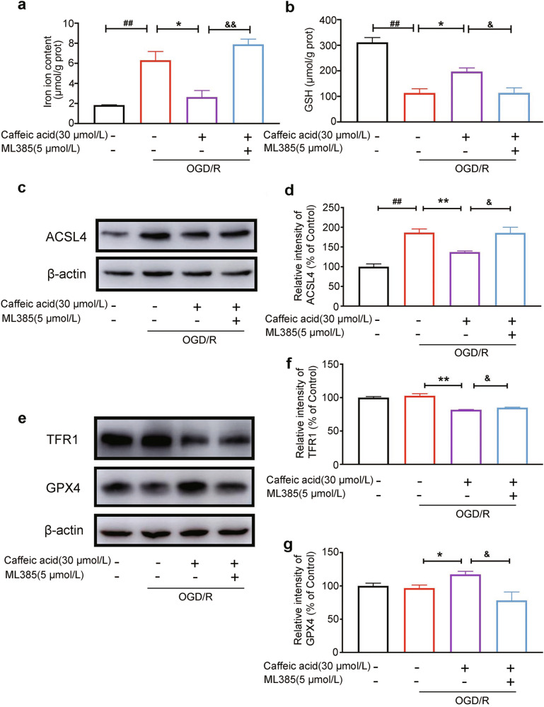
There are few effective and safe neuroprotective agents for the treatment of ischemic stroke currently. Caffeic acid is a phenolic acid that widely exists in a number of plant species. Previous studies show that caffeic acid ameliorates brain injury in rats after cerebral ischemia/reperfusion. In this study we explored the protective mechanisms of caffeic acid against oxidative stress and ferroptosis in permanent cerebral ischemia. Ischemia stroke was induced on rats by permanent middle cerebral artery occlusion (pMCAO). Caffeic acid (0.4, 2, 10 mg·kg-1·d-1, i.g.) was administered to the rats for 3 consecutive days before or after the surgery. We showed that either pre-pMCAO or post-pMCAO administration of caffeic acid (2 mg·kg-1·d-1) effectively reduced the infarct volume and improved neurological outcome. The therapeutic time window could last to 2 h after pMCAO. We found that caffeic acid administration significantly reduced oxidative damage as well as neuroinflammation, and enhanced antioxidant capacity in pMCAO rat brain. We further demonstrated that caffeic acid down-regulated TFR1 and ACSL4, and up-regulated glutathione production through Nrf2 signaling pathway to resist ferroptosis in pMCAO rat brain and in oxygen glucose deprivation/reoxygenation (OGD/R)-treated SK-N-SH cells in vitro. Application of ML385, an Nrf2 inhibitor, blocked the neuroprotective effects of caffeic acid in both in vivo and in vitro models, evidenced by excessive accumulation of iron ions and inactivation of the ferroptosis defense system. In conclusion, caffeic acid inhibits oxidative stress-mediated neuronal death in pMCAO rat brain by regulating ferroptosis via Nrf2 signaling pathway. Caffeic acid might serve as a potential treatment to relieve brain injury after cerebral ischemia. Caffeic acid significantly attenuated cerebral ischemic injury and resisted ferroptosis both in vivo and in vitro. The regulation of Nrf2 by caffeic acid initiated the transcription of downstream target genes, which were shown to be anti-inflammatory, antioxidative and antiferroptotic. The effects of caffeic acid on neuroinflammation and ferroptosis in cerebral ischemia were explored in a primary microglia-neuron coculture system. Caffeic acid played a role in reducing neuroinflammation and resisting ferroptosis through the Nrf2 signaling pathway, which further suggested that caffeic acid might be a potential therapeutic method for alleviating brain injury after cerebral ischemia.

Keywords: Nrf2; caffeic acid; ferroptosis; ischemic stroke; neuroinflammation; oxidative stress.


Conflict of interest statement

The authors declare no competing interests.

Figures

None
Caffeic acid significantly attenuated cerebral ischemic injury and resisted ferroptosis both in vivo and in vitro. The regulation of Nrf2 by caffeic acid initiated the transcription of downstream target genes, which were shown to be anti-inflammatory, antioxidative and antiferroptotic. The effects of caffeic acid on neuroinflammation and ferroptosis in cerebral ischemia were explored in a primary microglia-neuron coculture system. Caffeic acid played a role in reducing neuroinflammation and resisting ferroptosis through the Nrf2 signaling pathway, which further suggested that caffeic acid might be a potential therapeutic method for alleviating brain injury after cerebral ischemia.
Fig. 1
Fig. 1. Effects of caffeic acid on cerebral infarction volume, neurobehavioural scores, antioxidant enzymes and oxidative damage indices in pMCAO rats.
a Experimental design process. b, c Cerebral infarction volume of pMCAO rats. d mNSS Neurobehavioural scores of pMCAO rats. e Bederson Neurobehavioural scores of pMCAO rats (sham group: n = 3, vehicle and caffeic acid group: n = 15). f The levels of MDA in pMCAO rat brains (n = 5). g The levels of protein carbonylation in pMCAO rat brains (n = 5). h The level of 8-OHdG in pMCAO rat brains (n = 8). i The level of total antioxidant capacity in pMCAO rat brains (n = 10). j The activities of SOD in pMCAO rat brains (n = 13). k The activities of GSH-Px in pMCAO rat brains (n = 10). Data are shown as the mean ± SEM, #P < 0.05, ##P < 0.01, ###P < 0.001 vs. the sham group, *P < 0.05, **P < 0.01 and ***P < 0.001 vs. the vehicle group.
Fig. 2
Fig. 2. RNA-seq analysis of genes in pMCAO rat brain tissues.
a Genes in the sham group, vehicle group and caffeic acid group. b GO molecular function analysis in the vehicle group vs. the caffeic acid group. c Gene enrichment analysis (GSEA) indicated antioxidant- and anti-inflammation-related pathways, including flavin adenine dinucleotide binding, guanylate cyclase activity, oxygen carrier activity, and ATPase activity. (sham group: n = 3, vehicle group: n = 3 and caffeic acid group: n = 5).
Fig. 3
Fig. 3. Caffeic acid upregulates the Nrf2 signaling pathway after cerebral ischemia.
ac The mRNA transcription of the Nrf2 pathway in the brain tissue of pMCAO rats (n = 6–9). d, e Representative WB bands and the relative changes in p-AKT/AKT and p-GSK3β/GSK3β protein levels after caffeic acid treatment in pMCAO rats (n = 3). f, g Representative WB bands and the relative changes in Nrf2, HO-1, and NQO-1 protein levels after caffeic acid treatment in pMCAO rats (n = 3). h, i Representative WB bands and the relative changes in GCLC and GCLM protein levels after caffeic acid treatment in pMCAO rats (n = 3). Data are shown as the mean ± SEM, #P < 0.05, ##P < 0.01 and ###P < 0.001 vs. the sham group, *P < 0.05, **P < 0.01 and ***P < 0.001 vs. the vehicle group.
Fig. 4
Fig. 4. Caffeic acid exerted neuroprotective effects via the Nrf2 signaling pathway in OGD/R-treated SK-N-SH cells.
ad Representative WB bands and quantitative analysis of Nrf2, HO-1 and NQO-1 in the OGD/R-treated SK-N-SH cell model after caffeic acid treatment (n = 3). eh The effects of ML385 on the expression of Nrf2, HO-1, and NQO-1 (n = 3). i Cell viability was measured by MTT assay (n = 3). j MDA levels in OGD/R-damaged SK-N-SH cells treated with caffeic acid or ML385 (n = 3). k The level of total antioxidant capacity in OGD/R-damaged SK-N-SH cells treated with caffeic acid or ML385 (n = 3). l The level of GPx activity in OGD/R-damaged SK-N-SH cells treated with caffeic acid or ML385 (n = 4). The Western blot is representative of three independent experiments. Data are shown as the mean ± SEM, #P < 0.05, ##P < 0.01 and ###P < 0.001 vs. the control group, *P < 0.05, **P < 0.01 and ***P < 0.001 vs. the model group, &P < 0.05 and &&P < 0.01 vs. the caffeic acid group.
Fig. 5
Fig. 5. Caffeic acid inhibited ferroptosis in pMCAO rats.
a The protective effect of caffeic acid on intracellular iron content in pMCAO rats. (n = 5). b The improving effect of caffeic acid on GSH content in pMCAO rats (n = 5). ce The effects of caffeic acid on GSS and GSR protein levels (n = 3). f, j The effects of caffeic acid on GPX4 protein levels (n = 3). g, k The effects of caffeic acid on SLC3A2 protein levels (n = 3). h, l The effects of caffeic acid on ACSL4 protein levels (n = 3). i, m The effects of caffeic acid on TFR1 protein levels (n = 3). The Western blot is representative of three independent experiments. Data are shown as the mean ± SEM, #P < 0.05, ##P < 0.01, ###P < 0.001 vs. the sham group, *P < 0.05, **P < 0.01 and ***P < 0.001 vs. the vehicle group.
Fig. 6
Fig. 6. Caffeic acid inhibited ferroptosis in OGD/R-damaged cells.
a The protective effect of caffeic acid on intracellular iron content in OGD/R-treated SK-N-SH cells. (n = 3). b The improving effect of caffeic acid on GSH content in OGD/R-treated SK-N-SH cells (n = 3). c, d The effect of caffeic acid on GPX4 protein levels (n = 3). e, f The effect of caffeic acid on SLC3A2 protein levels (n = 3). g, h The effect of caffeic acid on ACSL4 protein levels (n = 3). i, j The effect of caffeic acid on TFR1 protein levels (n = 3). The Western blot is representative of three independent experiments. Data are shown as the mean ± SEM, #P < 0.05 and ###P < 0.001 vs. the control group, *P < 0.05, **P < 0.01 and ***P < 0.001 vs. the model group.
Fig. 7
Fig. 7. Caffeic acid reversed ferroptosis via the Nrf2 signaling pathway in vitro.
ML385, an inhibitor of Nrf2, reversed the protective effect of caffeic acid on intracellular iron content (a) and GSH content (b) in OGD/R-treated SK-N-SH cells (n = 3). The expression of ACSL4 (c, d), TFR1 (e, f) and GPX4 (e, g) was measured by Western blotting. The Western blot is representative of three independent experiments (n = 3). Data are shown as the mean ± SEM, ##P < 0.01 vs. the control group, *P < 0.05 and **P < 0.01 vs. the model group, &P < 0.05 and &&P < 0.01 vs. the caffeic acid group.
Fig. 8
Fig. 8. Caffeic acid reversed ferroptosis via the Nrf2 signaling pathway in vivo.
a, b Cerebral infarction volume of pMCAO rats. c mNSS Neurobehavioural scores of pMCAO rats. d Bederson Neurobehavioural scores of pMCAO rats (sham group: n = 3, vehicle group: n = 7, caffeic acid group: n = 7 and ML385 group: n = 6). ML385 reversed the protective effect of caffeic acid on iron content (e) and GSH content (f) in pMCAO rats (n = 3). g, i The effects of ML385 on the expression of Nrf2, HO-1, and NQO-1 (n = 3). h, j The effects of ML385 on the expression of ACSL4, TFR1, SLC7A11, and GPX4 (n = 3). Data are shown as the mean ± SEM, ##P < 0.01, ###P < 0.001 vs. the sham group, *P < 0.05, **P < 0.01 and ***P < 0.001 vs. the vehicle group, &P < 0.05, &&P < 0.01, and &&&P < 0.001 vs. the caffeic acid group.
Fig. 9
Fig. 9. Caffeic acid exerted anti-inflammatory properties in pMCAO rats.
a, b The expression of GFAP and COX2 was measured by Western blotting (n = 3). c, d Representative immunofluorescence staining of GFAP and the average fluorescence intensity (n = 3). Data are shown as the mean ± SEM, #P < 0.05, ##P < 0.01 vs. the sham group, *P < 0.05 and **P < 0.01 vs. the vehicle group.
Fig. 10
Fig. 10. Caffeic acid suppressed the LPS-induced proinflammatory response in BV2 cells via Nrf2 signaling.
The levels of TNF-α (a), IL-6 (b) and NO (c) were detected in the supernatant of LPS-stimulated BV2 cells (n = 4–5). Caffeic acid suppressed TNF-α (d), IL-6 (e), IL-1β (f) and NOS2 (g) mRNA levels in LPS-stimulated BV2 cells (n = 3–6). hj The effects of caffeic acid on Nfe2l2, Hmox1 and Nqo1 mRNA levels were measured by qRT‒PCR (n = 3–6). k, l Caffeic acid also suppressed the expression of iNOS in LPS-stimulated BV2 cells (n = 3). mq The effects of caffeic acid on Nrf2, HO-1 and NQO-1 expression were detected by Western blotting assays (n = 3). Data are shown as the mean ± SEM, ##P < 0.01 and ###P < 0.001 vs. the control group, *P < 0.05, **P < 0.01 and ***P < 0.001 vs. the model group.
Fig. 11
Fig. 11. Caffeic acid inhibited ferroptosis via the Nrf2 signaling pathway in OGD/R primary neurons and a coculture system of microglia and neurons.
a Experimental design process of OGD/R damage in primary neurons. b, c The effects of caffeic acid on Nrf2, HO-1 and NQO-1 protein levels in OGD/R primary neurons (n = 3). d, e The effects of caffeic acid on ACSL4, TFR1, SLC7A11 and GPX4 protein levels in OGD/R primary neurons (n = 3). f Experimental design process of OGD damage in a coculture system of microglia and neurons. The levels of TNF-α (g) and NO (h) were detected in the supernatant of LPS-stimulated primary microglia (n = 3). i, j The effects of caffeic acid on ACSL4, TFR1, SLC7A11 and GPX4 protein levels in primary neurons in the coculture system (n = 3). #P < 0.05, ##P < 0.01 and ###P < 0.001 vs. the control group, *P < 0.05, **P < 0.01 and ***P < 0.001 vs. the model group.

References

    1. Owolabi MO, Thrift AG, Mahal A, Ishida M, Martins S, Johnson WD, et al. Primary stroke prevention worldwide: translating evidence into action. Lancet Public Health. 2022;7:e74–e85. doi: 10.1016/S2468-2667(21)00230-9. - DOI - PMC - PubMed
    1. Cui Y, Zhang Y, Zhao X, Shao L, Liu G, Sun C, et al. Acsl4 exacerbates ischemic stroke by promoting ferroptosis-induced brain injury and neuroinflammation. Brain Behav Immun. 2021;93:312–21. doi: 10.1016/j.bbi.2021.01.003. - DOI - PubMed
    1. Cohen JE, Itshayek E, Moskovici S, Gomori JM, Fraifeld S, Eichel R, et al. State-of-the-art reperfusion strategies for acute ischemic stroke. J Clin Neurosci. 2011;18:319–23. doi: 10.1016/j.jocn.2010.10.008. - DOI - PubMed
    1. Kelmanson IV, Shokhina AG, Kotova DA, Pochechuev MS, Ivanova AD, Kostyuk AI, et al. In vivo dynamics of acidosis and oxidative stress in the acute phase of an ischemic stroke in a rodent model. Redox Biol. 2021;48:102178. doi: 10.1016/j.redox.2021.102178. - DOI - PMC - PubMed
    1. Sharpe PC, Mulholland C, Trinick T. Ascorbate and malondialdehyde in stroke patients. Ir J Med Sci. 1994;163:488–91. doi: 10.1007/BF02967089. - DOI - PubMed

MeSH terms

LinkOut - more resources