. 2024 Feb;47(1):264-284.
doi: 10.1007/s10753-023-01908-0. Epub 2023 Oct 14.

The Role of Nrf2 and Inflammation on the Dissimilar Cardiotoxicity of Doxorubicin in Two-Time Points: a Cardio-Oncology In Vivo Study Through Time

Affiliations

The Role of Nrf2 and Inflammation on the Dissimilar Cardiotoxicity of Doxorubicin in Two-Time Points: a Cardio-Oncology In Vivo Study Through Time

Ana Reis-Mendes et al. Inflammation. 2024 Feb.

Abstract

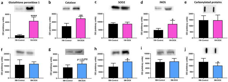
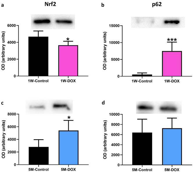
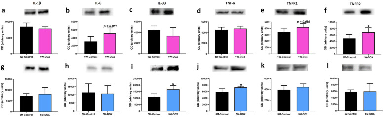
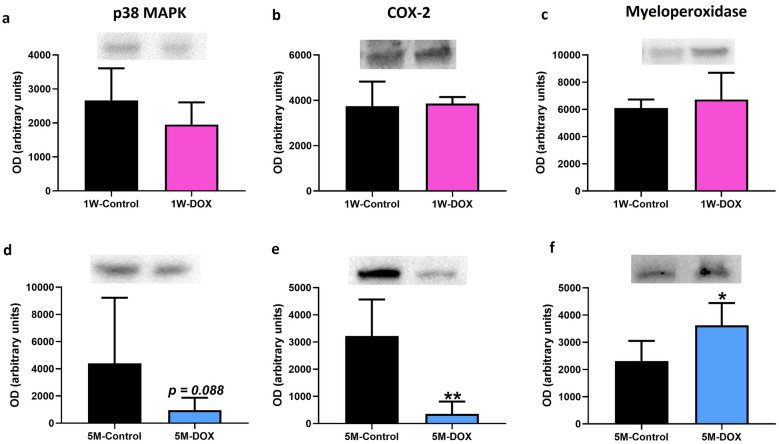
Doxorubicin (DOX) is a topoisomerase II inhibitor used in cancer therapy. Despite its efficacy, DOX causes serious adverse effects, such as short- and long-term cardiotoxicity. This work aimed to assess the short- and long-term cardiotoxicity of DOX and the role of inflammation and antioxidant defenses on that cardiotoxicity in a mice model. Adult CD-1 male mice received a cumulative dose of 9.0 mg/kg of DOX (2 biweekly intraperitoneal injections (ip), for 3 weeks). One week (1W) or 5 months (5M) after the last DOX administration, the heart was collected. One week after DOX, a significant increase in p62, tumor necrosis factor receptor (TNFR) 2, glutathione peroxidase 1, catalase, inducible nitric oxide synthase (iNOS) cardiac expression, and a trend towards an increase in interleukin (IL)-6, TNFR1, and B-cell lymphoma 2 associated X (Bax) expression was observed. Moreover, DOX induced a decrease on nuclear factor erythroid-2 related factor 2 (Nrf2) cardiac expression. In both 1W and 5M, DOX led to a high density of infiltrating M1 macrophages, but only the 1W-DOX group had a significantly higher number of nuclear factor κB (NF-κB) p65 immunopositive cells. As late effects (5M), an increase in Nrf2, myeloperoxidase, IL-33, tumor necrosis factor-α (TNF-α), superoxide dismutase 2 (SOD2) expression, and a trend towards increased catalase expression were observed. Moreover, B-cell lymphoma 2 (Bcl-2), cyclooxygenase-2 (COX-2), and carbonylated proteins expression decreased, and a trend towards decreased p38 mitogen-activated protein kinase (MAPK) expression were seen. Our study demonstrated that DOX induces adverse outcome pathways related to inflammation and oxidative stress, although activating different time-dependent response mechanisms.

Keywords: Nrf2.; cardiotoxicity; doxorubicin; inflammation; long-term effects; redox homeostasis disruption.


Conflict of interest statement

The authors declare no competing interests.

Figures

Fig. 1
Fig. 1
Body weight (a, b), water (c, d), and food (e, f) intake of mice sacrificed 1 week after the last administration of DOX (1W-DOX) and control mice (1W-Control); and mice sacrificed 5 months after the last administration of DOX (5M-DOX) and respective controls (5M-Control). DOX is shown in pink squares while controls are shown in black circles. Results are presented in grams (g) of body weight, mL of water intake/day/weight/animal, and g of food intake/day/weight/animal, as means ± SD. Statistical comparisons were made using a two-way ANOVA followed by Sidak’s post hoc test (*p < 0.05, **p < 0.01, ***p < 0.001, and ****p < 0.0001, DOX vs. control).
Fig. 2
Fig. 2
Representative light microscope micrographs were obtained after haematoxylin and eosin staining of the heart of mice, sacrificed 1 week after the last administration of DOX (1W-DOX) and control mice (1W-Control); and mice sacrificed 5 months after the last administration of DOX (5M-DOX) and respective controls (5M-Control), assessed by the haematoxylin and eosin staining. Control mice at 1W showed normal morphology and structure; mice given a cumulative dose of 9.0 mg/kg cumulative dose of DOX presented large and uncondensed nuclei (yellow arrow), interstitial oedema (white arrow), vacuolization (orange arrow), inflammatory infiltration (cyan arrow), and vascular congestion (green arrow). Necrotic zones (blue arrow) are evident. Scale bar = 100 µm, n = 3. Images were taken at 40 × magnification.
Fig. 3
Fig. 3
Representative light microscope micrographs were obtained after Sirius red staining of the heart of mice sacrificed one week after the last administration of DOX (1W-DOX) and control mice (1W-Control), and mice sacrificed 5 months after the last administration of DOX (5M-DOX) and respective controls (5M-Control). Scale bar = 100 µm, n = 3 animals per group, six random fields per animal. Images were taken at 40 × magnification. ab The results of collagen content red staining are expressed as a percentage of collagen per total section area and are presented as means ± SD. Statistical comparisons were made using the Mann–Whitney test: *p < 0.05, DOX vs. control.
Fig. 4
Fig. 4
Western blotting analysis of a, f glutathione peroxidase (22 kDa), b, g catalase (60 kDa), c, h superoxide dismutase 2 (SOD2) (26.6 kDa) and d, i inducible nitric oxide synthase (iNOS) (131 kDa) expression in the cardiac tissue. e, j Protein carbonylation cardiac content was evaluated by slot blot. a, b, c, d, e Mice sacrificed 1 week after the last administration of DOX (1W-DOX) and control mice (1W-Control); and f, g, h, i, j mice sacrificed 5 months after the last administration of DOX (5M-DOX) and respective controls (5M-Control). Values are expressed as mean ± SD and were obtained from 4–6 (1W) to 4–7 (5M) animals from each treatment group. Statistical comparisons were made using the unpaired t-test: *p < 0.05, **p < 0.01, ****p < 0.0001, DOX vs. control. OD, optic density. Protein loading was confirmed by the Ponceau S staining (Fig. S1).
Fig. 5
Fig. 5
Western blotting analysis of a, c nuclear factor erythroid-2 related factor 2 (Nrf2) (97 kDa) and c, d p62 (62 kDa) expression in the cardiac tissue, in a, b mice sacrificed 1 week after the last administration of DOX (1W-DOX) and control mice (1W-Control); and c, d mice sacrificed 5 months after the last administration of DOX (5M-DOX) and respective controls (5M-Control). Values are expressed as mean ± SD and were obtained from 4–6 (1W) to 5–6 (5M) animals from each treatment group. Statistical comparisons were made using the unpaired t-test: *p < 0.05, ***p < 0.001, DOX vs. control. OD, optic density. Protein loading was confirmed by the Ponceau S staining (Fig. S2).
Fig. 6
Fig. 6
a Representative images of the immunohistochemistry determination of macrophages markers CD68 (a marker for the macrophage M1) (on the top line) and b CD206 (a marker for the macrophage M2) (the bottom histologic figures) in cardiac tissue of mice sacrificed 1 week after the last administration of DOX (1W-DOX) and control mice (1W-Control). c, d The number of cells staining as positive, indicated by yellow arrows for the activated macrophages marked as M1 of the heart of DOX-treated and control groups, c 1W-DOX and 1W-Control; and d 5M-DOX and respective 5M-Control. e, f The number of cells staining as positive, indicated by yellow arrows for the activated macrophages marked as M2 of the heart of DOX-treated and control groups, e 1W-DOX and 1W-Control; and f 5M-DOX and respective 5M-Control. The results were expressed according to the number of positive cells per area (µm2) as mean ± SD. Statistical comparisons were made using the Mann–Whitney test: **p < 0.01, ***p < 0.001, DOX vs. control. Scale bar = 100 µm, n = 3 animals per group, six random fields per animal. Images were taken at 40× magnification.
Fig. 7
Fig. 7
Western blotting analysis of a, g interleukin-1β (IL-1 β) (35 kDa), b, h interleukin-6 (IL-6) (23 kDa), c, i interleukin-33 (IL-33) (33 kDa), d, j tumor necrosis factor-α (TNF-α) (25 kDa), e, k type 1 TNF receptor (TNFR1) (50 kDa) and f, l type 2 TNF receptor (TNFR2) (75 kDa) expression in the cardiac tissue, in a, b, c mice sacrificed 1 week after the last administration of DOX (1W-DOX) and control mice (1W-Control); and d, e, f mice sacrificed 5 months after the last administration of DOX (5M-DOX) and respective controls (5M-Control). Values are expressed as mean ± SD and were obtained from 4–6 (1W) to 4–6 (5M) animals from each treatment group. Statistical comparisons were made using the unpaired t-test: *p < 0.05, DOX vs. control. OD, optic density. Protein loading was confirmed by the Ponceau S staining (Fig. S3).
Fig. 8
Fig. 8
Western blotting analysis of a, d p38 mitogen-activated protein kinase (p38 MAPK) (40 kDa), b, e cyclooxygenase-2 (COX-2) (75 kDa), c, f myeloperoxidase (63 kDa) expression in the cardiac tissue, in a, b, c mice sacrificed 1 week after the last administration of DOX (1W-DOX) and control mice (1W-Control); and d, e, f mice sacrificed 5 months after the last administration of DOX (5M-DOX) and respective controls (5M-Control). Values are expressed as mean ± SD and were obtained from 4–6 (1W) to 5–7 (5M) animals from each treatment group. Statistical comparisons were made using the unpaired t-test: *p < 0.05, **p < 0.01, DOX vs. control. OD, optic density. Protein loading was confirmed by the Ponceau S staining (Fig. S4).
Fig. 9
Fig. 9
Representative images of the immunohistochemistry determination of nuclear factor κB (NF-κB) in the cardiomyocytes-like cells from mice sacrificed 1 week after the last administration of DOX (1W-DOX) and control mice (1W-Control), and mice sacrificed 5 months after the last administration of DOX (5M-DOX) and respective controls (5M-Control). a, c The number of cells staining as positive, indicated by black arrows for the activated NF-κB of the heart of DOX-treated and control groups, a 1W-DOX and 1W-Control; and c 5M-DOX and respective 5M-Control. Scale bar = 100 µm, n = 3 animals per group, six random fields per animal. Images were taken at 40 × magnification. The results were expressed according to the number of positive cells per area (µm2) as mean ± SD. Statistical comparisons were made using the Mann–Whitney test: *p < 0.05, DOX vs. control. b, d Western blotting analysis of NF-κB p65 (60 kDa) expression in the cardiac tissue, in b 1W-DOX and 1W-Control; and d 5M-DOX and 5M-Control. Values are expressed as mean ± SD and were obtained from 6 (1W) to 7 (5M) animals from each treatment group. Statistical comparisons were made using the unpaired t-test. OD, optic density. Protein loading on the Western blot was confirmed by the Ponceau S staining (Fig. S5).
Fig. 10
Fig. 10
Western blotting analysis of a, d B-cell lymphoma 2 (Bcl-2) (26 kDa) and b, e B-cell lymphoma 2 associated X (Bax) (21 kDa) and c, f ratio of Bax/Bcl-2 expression in the cardiac tissue, in a, b, c mice sacrificed 1 week after the last administration of DOX (1W-DOX) and control mice (1W-Control); and d, e, f mice sacrificed 5 months after the last administration of DOX (5M-DOX) and respective controls (5M-Control). Values are expressed as mean ± SD and were obtained from 4–6 (1W) to 4–7 (5M) animals from each treatment group. Statistical comparisons were made using the unpaired t-test: *p < 0.05, DOX vs. control. OD, optic density. Protein loading was confirmed by the Ponceau S staining (Fig. S6).

References

    1. Reis-Mendes A, Sousa E, de Lourdes Bastos M, Costa VM. The role of the metabolism of anticancer drugs in their induced-cardiotoxicity. Current Drug Metabolism. 2015 doi: 10.2174/1389200216666151103114926. - DOI - PubMed
    1. Nebigil CG, Désaubry L. Updates in anthracycline-mediated cardiotoxicity. Frontiers in Pharmacology. 2018 doi: 10.3389/fphar.2018.01262. - DOI - PMC - PubMed
    1. Renu K, Abilash VG, Tirupathi Pichiah PB, Arunachalam S. Molecular mechanism of doxorubicin-induced cardiomyopathy - An update. European Journal of Pharmacology. 2018 doi: 10.1016/j.ejphar.2017.10.043. - DOI - PubMed
    1. Todorova VK, Wei JY, Makhoul I. Subclinical doxorubicin-induced cardiotoxicity update: role of neutrophils and endothelium. American Journal of Cancer Research. 2021;11(9):4070–4091. - PMC - PubMed
    1. Wallace KB. Doxorubicin-induced cardiac mitochondrionopathy. Pharmacology and Toxicology. 2003 doi: 10.1034/j.1600-0773.2003.930301.x. - DOI - PubMed