. 2023 Oct 13;22(1):276.
doi: 10.1186/s12933-023-02010-3.

Direct regulation of the cardiac ryanodine receptor (RyR2) by O-GlcNAcylation

Affiliations

Direct regulation of the cardiac ryanodine receptor (RyR2) by O-GlcNAcylation

Chidinma A Okolo et al. Cardiovasc Diabetol. .

Abstract

Background: O-GlcNAcylation is the enzymatic addition of a sugar, O-linked β-N-Acetylglucosamine, to the serine and threonine residues of proteins, and is abundant in diabetic conditions. We have previously shown that O-GlcNAcylation can trigger arrhythmias by indirectly increasing pathological Ca2+ leak through the cardiac ryanodine receptor (RyR2) via Ca2+/calmodulin-dependent kinase II (CaMKII). However, RyR2 is well known to be directly regulated by other forms of serine and threonine modification, therefore, this study aimed to determine whether RyR2 is directly modified by O-GlcNAcylation and if this also alters the function of RyR2 and Ca2+ leak.

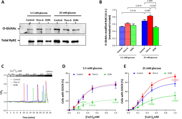
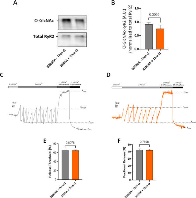
Methods: O-GlcNAcylation of RyR2 in diabetic human and animal hearts was determined using western blotting. O-GlcNAcylation of RyR2 was pharmacologically controlled and the propensity for Ca2+ leak was determined using single cell imaging. The site of O-GlcNAcylation within RyR2 was determined using site-directed mutagenesis of RyR2.

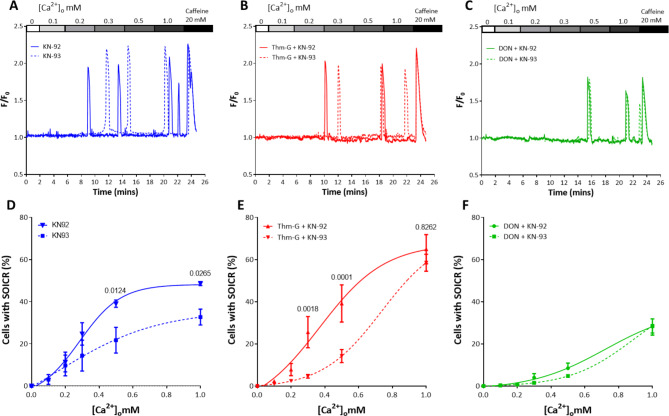
Results: We found that RyR2 is modified by O-GlcNAcylation in human, animal and HEK293 cell models. Under hyperglycaemic conditions O-GlcNAcylation was associated with an increase in Ca2+ leak through RyR2 which persisted after CaMKII inhibition. Conversion of serine-2808 to alanine prevented an O-GlcNAcylation induced increase in Ca2+ leak.

Conclusions: These data suggest that the function of RyR2 can be directly regulated by O-GlcNAcylation and requires the presence of serine-2808.

Keywords: Diabetes; O-GlcNAcylation; Ryanodine receptor (RyR2); SOICR.


Conflict of interest statement

The authors declare no competing interests.

Figures

Fig. 1
Fig. 1
RyR2 is O-GlcNAcylated. (A) Representative blots showing total O-GlcNAc-RyR2 and total RyR2 expression from diabetic and non-diabetic human right atrial appendages (RAA) and left ventricles of Zucker diabetic fatty (ZDF) rat and non-diabetic lean (control) rats. The samples are labelled above the corresponding lane. O-GlcNAc modified RyR2 normalised to total RyR2 expression in (B) human RAA samples (n = 8 per group) and, (C) rat LV samples (n = 9 per group). Data displayed as Mean ± SEM. Statistical analysis was performed using a Welch’s t test
Fig. 2
Fig. 2
Changes in RyR2 O-GlcNAcylation are associated with changes in SOICR. Application of Thm-G and DON on the O-GlcNAc-RyR2 level in cells grown in low glucose (5.5 mM) and high glucose (25 mM) media. (A) Representative blots showing O-GlcNAc-RyR2 and total RyR2. (B) Densitometric analysis of O-GlcNAc-RyR2 level. All O-GlcNAc-RyR2 was normalised to total RyR2. Results are shown as Mean ± SEM. n = 3 independent lysates. Statistical analysis was performed using two-way ANOVA (Šidák post-hoc test). (C) Representative Fluo-4 traces presented as F/F0. Peaks represent SOICR events. (D) Percentage of cells experiencing SOICR at 5.5 mM glucose, (E) percentage of cells experiencing SOICR at 25 mM glucose. Data displayed are the average of 5–8 independent experiments and expressed as Mean ± SEM. Only significant P-values, vs. control are shown. Statistical analysis was performed using two-way ANOVA (Tukey post-hoc test)
Fig. 3
Fig. 3
CaMKII inhibition does not prevent an O-GlcNAc mediated increase SOICR propensity. (A-C) Representative Fluo-4 traces (F/F0) of cells treated with KN-92 or KN-93 in the absence (A) or presence of Thm-G (B) or DON (C). Peaks represent SOICR events. (D-F) Percentage of cells experiencing SOICR treated with KN-92 or KN-93 in the absence (D) or presence of Thm-G (E) or DON (F). Data displayed are the average of 4–12 independent experiments and expressed as Mean ± SEM. P-values shown are for comparison of cells treated with KN-92 vs. KN-93. Statistical analysis was performed using two-way ANOVA (with Šidák post-hoc test)
Fig. 4
Fig. 4
Effect of Thm-G and CaMKII inhibition on the characteristics of SOICR. Representative CEPIA traces for (A) KN-92 treated cells, cells treated with (B) KN-93, (C) Thm-G + KN-92 and (D) Thm-G + KN-93. Scale bar shows fluorescence (10AU) vs. time (60s). (E) Comparison of FSOICR, and (F) fractional release between treatments. Data were obtained from 156–542 cells recorded over 6–14 independent experiments. Results are expressed as Mean ± SEM. Statistical analysis was performed using a two-way ANOVA (with Fisher’s LSD post-hoc test)
Fig. 5
Fig. 5
S2808 is required for CaMKII independent O-GlcNAcylation mediated changes in SOICR. (A) Representative blots showing O-GlcNAc-RyR2 and total RyR2. (B) Densitometric analysis of O-GlcNAc-RyR2 level. All O-GlcNAc-RyR2 was normalised to total RyR2. Results are shown as Mean ± SEM. n = 4 independent lysates. Statistical analysis was performed using an unpaired t-test. (C) Representative CEPIA traces for S2808 cells without Thm-G (A) and with Thm-G treatment (D). Comparison of (E) release threshold, and (F) fractional release in S2808 cells treated with or without Thm-G. Results were obtained from 125 and 135 cells (for – and + Thm-G, respectively) recorded over 8 independent experiments. To remove the impact of CaMKII, KN-93 was present in all conditions. Results are expressed as Mean ± SEM. Statistical analysis was performed using a Mann-Whitney test

References

    1. Bers DM. Cardiac excitation-contraction coupling. Nature. 2002;415:198–205. doi: 10.1038/415198a. - DOI - PubMed
    1. Zhang JZ, McLay JC, Jones PP. The arrhythmogenic human HRC point mutation S96A leads to spontaneous ca release due to an impaired ability to buffer store ca. J Mol Cell Cardiol. 2014;74C:22–31. doi: 10.1016/j.yjmcc.2014.04.019. - DOI - PubMed
    1. Jones PP, Guo W, Chen SRW. Control of cardiac ryanodine receptor by sarcoplasmic reticulum luminal Ca2. J Gen Physiol. 2017;149(9):867–75. doi: 10.1085/jgp.201711805. - DOI - PMC - PubMed
    1. Dobrev D, Wehrens XH. Role of RyR2 phosphorylation in heart failure and arrhythmias: Controversies around ryanodine receptor phosphorylation in cardiac disease Circ Res, 2014. 114(8): p. 1311-9; discussion 1319. - PMC - PubMed
    1. Gonano LA, et al. Regulation of cardiac ryanodine receptor function by the cyclic-GMP dependent protein kinase G. Curr Res Physiol. 2022;5:171–8. doi: 10.1016/j.crphys.2022.03.004. - DOI - PMC - PubMed

Publication types

MeSH terms