. 2023 Oct 13;43(1):49.
doi: 10.1186/s41232-023-00300-7.

Testosterone upregulates glial cell line-derived neurotrophic factor (GDNF) and promotes neuroinflammation to enhance glioma cell survival and proliferation

Affiliations

Testosterone upregulates glial cell line-derived neurotrophic factor (GDNF) and promotes neuroinflammation to enhance glioma cell survival and proliferation

Kouminin Kanwore et al. Inflamm Regen. .

Abstract

Background: Testosterone contributes to male organism development, such as bone density, muscle development, and fat repartition. Estrogen (derived from testosterone) also contributes to female reproductive system development. Here, we investigated the effect of testosterone on glioma cells and brain neuron inflammation essential for cancer development and progression.

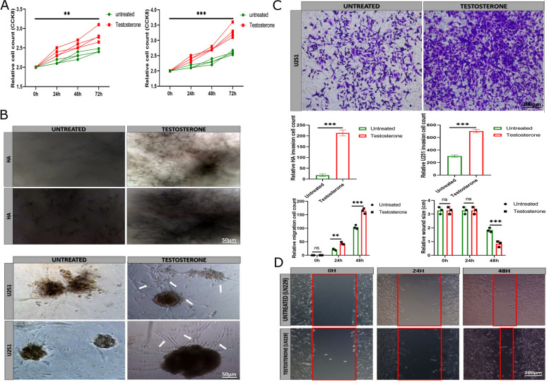
Methods: The human astrocyte and glioma cell lines were treated with 6 ng/ml exogenous testosterone in vitro. We performed cell counting kit-8, transwell, and wound healing assays to determine the effect of testosterone on glioma cell proliferation, migration, and invasion. The glioma cells were injected into the xenograft and treated with 5 µl concentrated testosterone. Transcriptional suppression of glial cell line-derived neurotrophic factor (GDNF) was performed to evaluate brain neuron inflammation and survival. The tumor tissues were assessed by hematoxylin-eosin staining and immunohistochemistry.

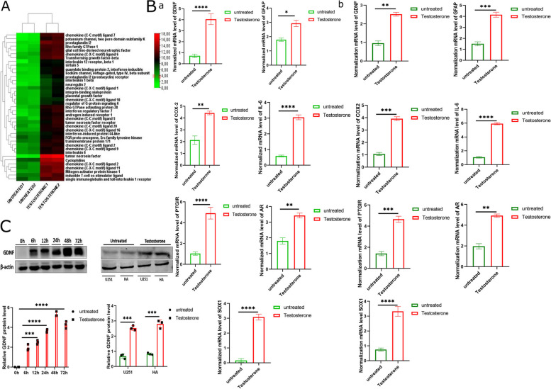
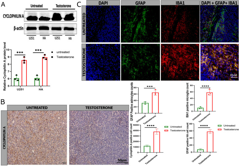
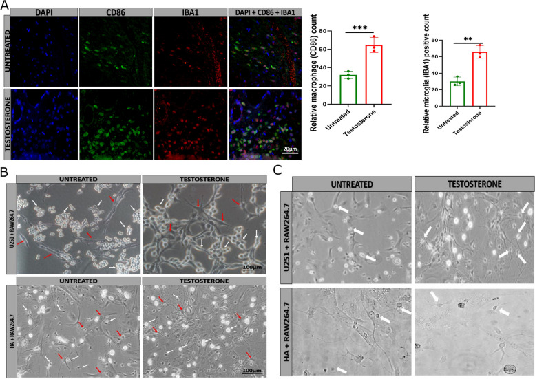
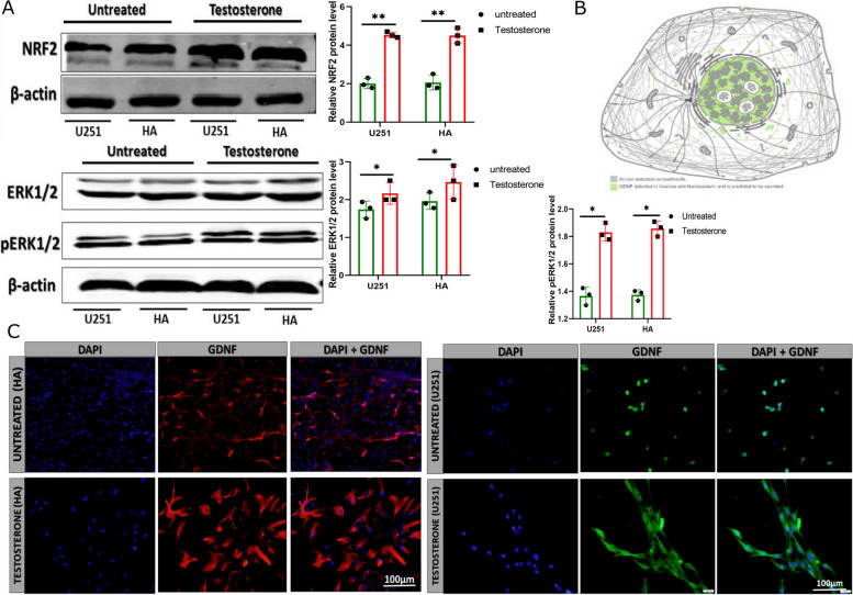
Results: Testosterone upregulates GDNF to stimulate proliferation, migration, and invasion of glioma cells. Pathologically, the augmentation of GDNF and cyclophilin A contributed to neuroprotection when treated with testosterone. Our investigation showed that testosterone contributes to brain neuron and astrocyte inflammation through the upregulation of nuclear factor erythroid 2-related factor 2 (NRF2), glial fibrillary acid protein (GFAP), and sirtuin 5 (SIRT5), resulting in pro-inflammatory macrophages recruitments into the neural microenvironment. Mechanically, testosterone treatment regulates GDNF translocation from the glioma cells and astrocyte nuclei to the cytoplasm.

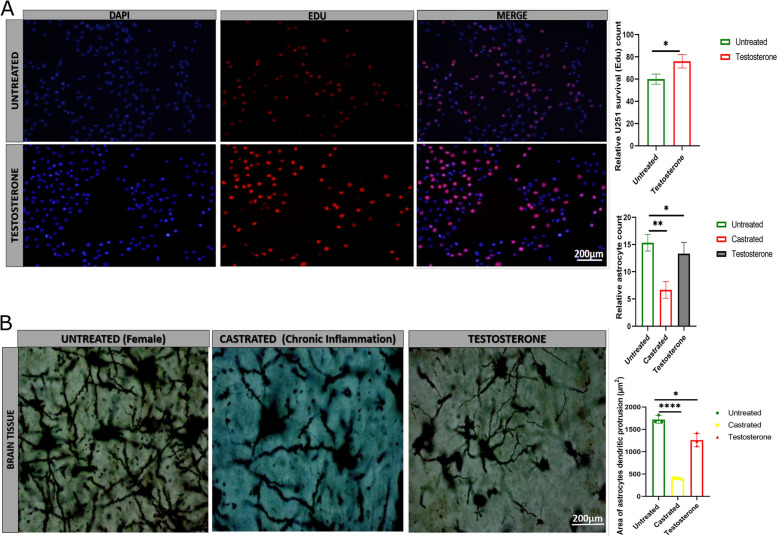
Conclusion: Testosterone upregulates GDNF in glioma cells and astrocytes essential for microglial proliferation, migration, and invasion. Testosterone contributes to brain tumor growth via GDNF and inflammation. The contribution of testosterone, macrophages, and astrocytes, in old neuron rescue, survival, and proliferation. During brain neuron inflammation, the organism activates and stimulates the neuron rescue through the enrichment of the old neuron microenvironment with growth factors such as GDNF, BDNF, SOX1/2, and MAPK secreted by the surrounding neurons and glial cells to maintain the damaged neuron by inflammation alive even if the axon is dead. The immune response also contributes to brain cell survival through the secretion of proinflammatory cytokines, resulting in inflammation maintenance. The rescued old neuron interaction with infiltrated macrophages contributes to angiogenesis to supplement the old neuron with more nutrients leading to metabolism activation and surrounding cell uncontrollable cell growth.

Keywords: Cyclophilin A; GDNF; Glioma; Neuroinflammation; Neuroprotection; Testosterone.


Conflict of interest statement

The authors declare that they have no competing interests.

Figures

Fig. 1
Fig. 1
Differential gene expression between the control (untreated) and testosterone-treated U251 and HA. A Next-generation sequencing showing differential gene expression between the untreated group and testosterone-treated U251 (n = 2). We selected the genes that are involved in neuroinflammation only. B Reverse transcription-quantitative polymerization chain reaction (RT-qPCR) comparing GDNF, SOX1, IL-6, COX2, PTGIR, AR, and GFAP between the control (untreated) and testosterone-treated U251 and HA, a for U251 and b for HA (n = 3). C Immunoblotting comparing GDNF protein levels between the control (untreated) and testosterone-treated U251 and HA (n = 3). *p < 0.05, **p < 0.01, ***p < 0.001, and ****p < 0.0001
Fig. 2
Fig. 2
The effect of testosterone treatment on U251 and HA proliferation, invasion, and migration. A Cell counting kit 8 indicating U251 and HA survival and proliferation (n = 4). B Invasion assay comparing the untreated and testosterone-treated U251 and HA invasion on agarose gel (n = 3). The white arrow indicates invasive U251. C Transwell matrigel assay comparing the untreated and testosterone-treated U251 invasion and migration (n = 3). D wound-healing assay comparing the untreated and testosterone-treated LN229 migration (n = 3). **p < 0.01, ***p < 0.001, and ****p < 0.0001
Fig. 3
Fig. 3
Testosterone upregulates cyclophilin A expression in vitro and in vivo. A Immunoblotting detection of cyclophilin A protein level in U251 and HA (n = 3). B Immunohistochemistry comparing cyclophilin A-positive cells between the untreated and testosterone-treated xenograft tissue (n = 3). C Immunofluorescence comparing GFAP (green) and IBA1 (red) positive inflammatory astrocyte between the untreated and testosterone-treated brain tissue (n = 3). GFAP (green) indicated the total astrocyte and IBA1 of the inflammatory microglia. ***p < 0.001, and ****p < 0.0001
Fig. 4
Fig. 4
Testosterone contributes to U251 and HA survival. A 5-Ethynyl-2’-Deoxyuridine (EDU) staining comparing U251 survival between the untreated and testosterone-treated groups (n = 3). B Comparison of brain astrocyte shape (protrusions) between the untreated and testosterone-treated brain tissue in inflammation condition using Sholl analysis (n = 3). In the normal female mouse tissue, the astrocytes' shape looked good, and the axons and dendritic protrusions were well differentiated. In the castrated mouse tissue, the astrocyte shape and axons are affected (altered and discontinuous axon protrusions) due to tumor inflammation and a decrease in testosterone level. In testosterone-treated castrated mice, we can observe the regeneration of axon protrusions and improvement of the astrocyte shape even under inflammation. *p < 0.05 and **p < 0.01
Fig. 5
Fig. 5
Testosterone promotes pro-inflammatory macrophage recruitment and activation. A Immunofluorescence comparing pro-inflammatory macrophages (CD86) and microglial (Iba1) between the untreated and testosterone-treated tumor tissue (n = 3) B Contrast image showing macrophage (Raw264.7) interaction with U251 and HA between the untreated and testosterone-treated groups (n = 4). The white arrow indicates the macrophages and the red arrow indicates U251 or HA. C Contrast image comparing macrophage activation between the untreated and testosterone-treated groups (n = 4). The activated microphage have outgrowth while the non-activated macrophages did not have. **p < 0.01 and ***p < 0.001
Fig. 6
Fig. 6
The testosterone upregulates pro-inflammatory genes NRF2, ERK1/2, and pERK1/2 protein level and GDNF translocation. A Immunoblotting detection of NRF2, ERK1/2, and pERK1/2 protein levels in U251 and HA (n = 3). B Protein atlas indicating GDNF subcellular location. C Immunofluorescence detection of GDNF subcellular localization in human astrocyte (red) and U251 (green) (n = 4). *p < 0.05 and **p < 0.01
Fig. 7
Fig. 7
The effect of GDNF knockout on inflammatory protein level, pro-inflammatory macrophage recruitment, and microglial survival. A Western blot detection of NRF2, iNOS, ERK1/2, pERK1/2, CD40L, GDNF, and Survivin in castrated, testosterone, female, and GDNF knockout (n = 3). B Immunofluorescence detection of pro-inflammatory macrophages (CD86) and microglia (Iba1) between the untreated and GDNF knockout + testosterone-treated brain tissue, Dapi was used to stain U251 nuclei (n = 3). C Edu staining comparing the untreated and GDNF knockout + testosterone treated U251 survival (n = 3). Dapi was used to stain U251 nuclei. *p < 0.05, **p < 0.01, ***p < 0.001, ****p < 0.0001

References

    1. Carrano A, Juarez JJ, Incontri D, Ibarra A, Guerrero Cazares H. Sex-specific differences in glioblastoma. Cells. 2021;10(7). 10.3390/cells10071783. - PMC - PubMed
    1. Ostrom QT, Kinnersley B, Wrensch MR, Eckel-Passow JE, Armstrong G, Rice T, et al. Sex-specific glioma genome-wide association study identifies new risk locus at 3p21.31 in females, and finds sex-differences in risk at 8q24.21. Sci Rep. 2018;8(1):7352. - PMC - PubMed
    1. Vining B, Ming Z, Bagheri-Fam S, Harley V. Diverse regulation but conserved function: SOX9 in vertebrate sex determination. Genes (Basel). 2021;12(4). 10.3390/genes12040486. - PMC - PubMed
    1. Jakob S, Lovell-Badge R. Sex determination and the control of Sox9 expression in mammals. FEBS J. 2011;278(7):1002–1009. - PubMed
    1. Garcia I, Aldaregia J, MarjanovicVicentic J, Aldaz P, Moreno-Cugnon L, Torres-Bayona S, et al. Oncogenic activity of SOX1 in glioblastoma. Sci Rep. 2017;7:46575. - PMC - PubMed

LinkOut - more resources