. 2023 Sep 22;24(19):14420.
doi: 10.3390/ijms241914420.

Enhanced Antibacterial Ability of Electrospun PCL Scaffolds Incorporating ZnO Nanowires

Affiliations

Enhanced Antibacterial Ability of Electrospun PCL Scaffolds Incorporating ZnO Nanowires

Jingjing Tian et al. Int J Mol Sci. .

Abstract

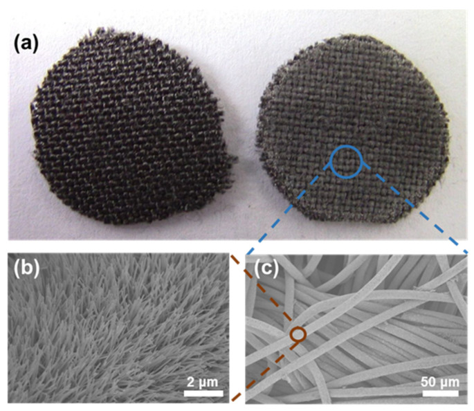
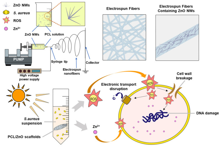
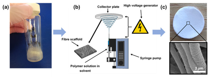
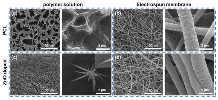
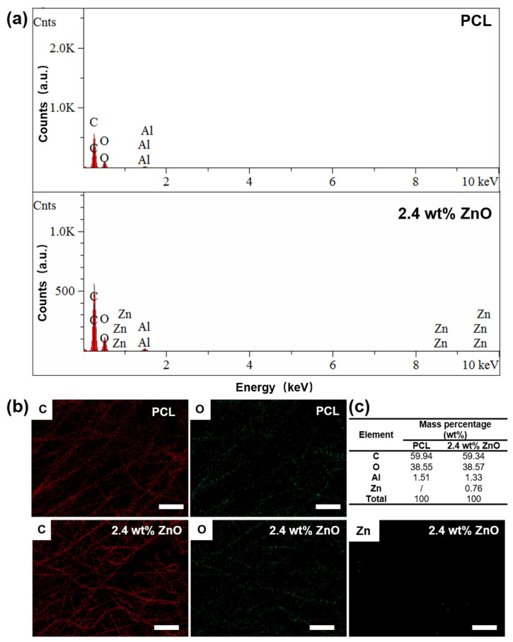
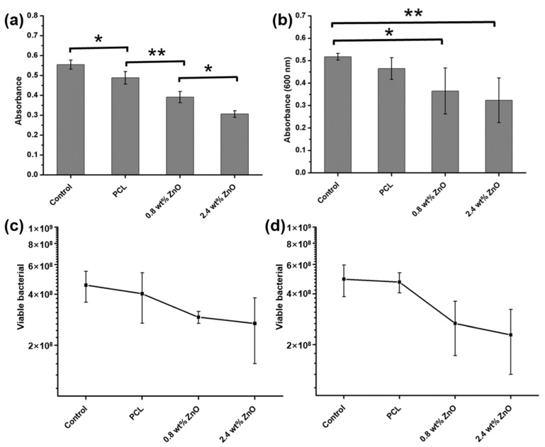
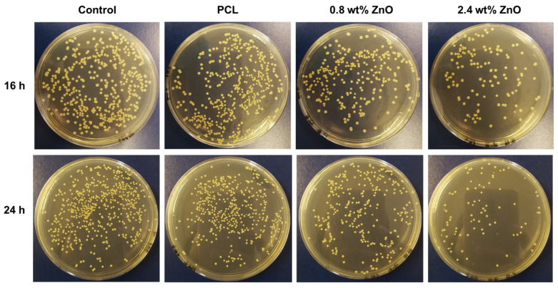
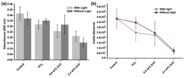
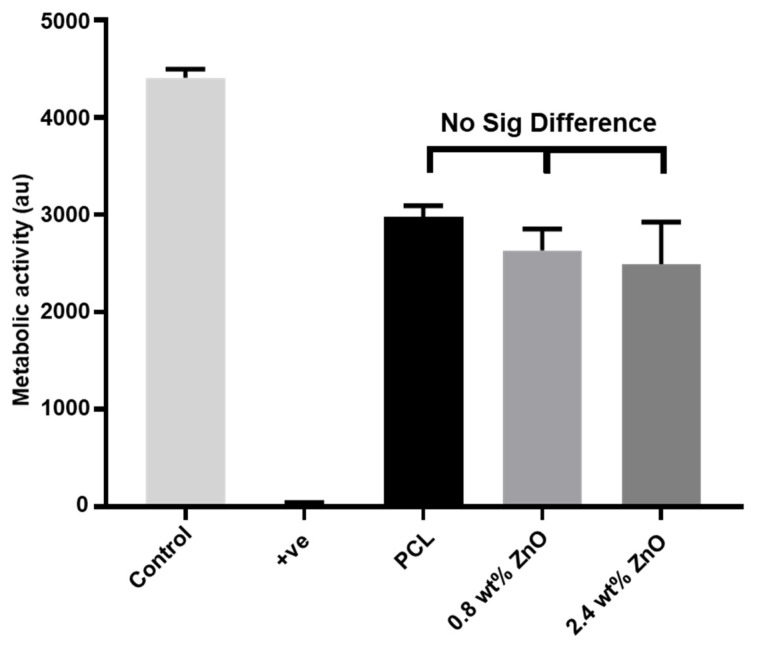
The infection of implanted biomaterial scaffolds presents a major challenge. Existing therapeutic solutions, such as antibiotic treatment and silver nanoparticle-containing scaffolds are becoming increasingly impractical because of the growth of antibiotic resistance and the toxicity of silver nanoparticles. We present here a novel concept to overcome these limitations, an electrospun polycaprolactone (PCL) scaffold functionalised with zinc oxide nanowires (ZnO NWs). This study assessed the antibacterial capabilities and biocompatibility of PCL/ZnO scaffolds. The fabricated scaffolds were characterised by SEM and EDX, which showed that the ZnO NWs were successfully incorporated and distributed in the electrospun PCL scaffolds. The antibacterial properties were investigated by co-culturing PCL/ZnO scaffolds with Staphylococcus aureus. Bacterial colonisation was reduced to 51.3% compared to a PCL-only scaffold. The biocompatibility of the PCL/ZnO scaffolds was assessed by culturing them with HaCaT cells. The PCL scaffolds exhibited no changes in cell metabolic activity with the addition of the ZnO nanowires. The antibacterial and biocompatibility properties make PCL/ZnO a good choice for implanted scaffolds, and this work lays a foundation for ZnO NWs-infused PCL scaffolds in the potential clinical application of tissue engineering.

Keywords: PCL scaffold; ZnO nanowire; antimicrobial; electrospinning; tissue engineering.


Conflict of interest statement

The authors declare no conflict of interest.

Figures

Figure 1
Figure 1
Fabrication and characterisation of the ZnO NWs. (a) Carbon cloth with a diameter of 10 mm before (left) and after (right) ZnO NWs growth. (b) ZnO NWs grown on one fibre of carbon cloth imaged using SEM. (c) ZnO NWs grown along the textile fibres of carbon cloth.
Scheme 1
Scheme 1
Schematic diagram of the preparing of PCL/ZnO scaffolds using electrospinning and the underlying antimicrobial mechanism.
Figure 2
Figure 2
Preparation of electrospun PCL/ZnO scaffolds. (a) The ZnO NWs were dissolved in PCL suspension to obtain a thorough mixture. (b) Experimental setup for electrospinning. (c) Electrospun PCL/ZnO nanofiber membranes and magnified SEM image of the fibres.
Figure 3
Figure 3
Characterisation of the electrospinning suspension and PCL/ZnO scaffolds using SEM. (a) SEM image of the PCL polymer suspension. (b) SEM image of the electrospun PCL membrane. (c) SEM image of PCL polymer suspension containing ZnO NWs. (d) The SEM image of the PCL/ZnO scaffolds.
Figure 4
Figure 4
EDX Characterisation of PCL/ZnO scaffold. (a) The EDX spectrum image of PCL and 2.4% wt% PCL/ZnO scaffolds. (b) C (red), O (green) and Zn element (orange) analysis of PCL and 2.4% wt% PCL/ZnO scaffolds using EDX. (c) The mass percentage of PCL and 2.4% wt% PCL/ZnO scaffolds measured using EDX. Scar bar is 10 μm.
Figure 5
Figure 5
ZnO NWs dose-dependently sterilised PCL/ZnO scaffolds against S. aureus. (a) Absorbance of bacterial suspension at 600 nm after co-culture with various scaffolds for 16 h. (b) Absorbance of bacterial suspension after co-culture with various scaffolds for 24 h. (c) Viable bacteria in the bacterial suspension after co-culture with various scaffolds for 16 h detected using the colony counting method. (d) Viable bacteria in the bacterial suspension after co-culture with various scaffolds for 24 h detected using the colony counting method. * p < 0.05, ** p < 0.01.
Figure 6
Figure 6
Bacterial colonies after S. aureus came directly into contact with PCL, 0.8 wt% and 2.4 wt% PCL/ZnO scaffolds for 16 and 24 h, respectively.
Figure 7
Figure 7
Effect of visible-light irradiation on the antibacterial properties of PCL/ZnO scaffolds. (a) Absorbance of bacterial suspension with/without visible light irradiation after co-culture with various scaffolds for 40 h. (b) Viable bacteria in suspension with/without visible light irradiation after co-culture with various scaffolds for 40 h detected using the colony counting method.
Figure 8
Figure 8
The metabolic activity of the PCL/ZnO scaffold measured using PrestoBlueTM assay after co-culture with HaCaT cells for 3 days.

References

    1. Siddiqui N., Asawa S., Birru B., Baadhe R., Rao S. PCL-Based Composite Scaffold Matrices for Tissue Engineering Applications. Mol. Biotechnol. 2018;60:506–532. doi: 10.1007/s12033-018-0084-5. - DOI - PubMed
    1. Qian Y., Zhou X., Zhang F., Diekwisch T.G., Luan X., Yang J. Triple PLGA/PCL Scaffold Modification Including Silver Impregnation, Collagen Coating, and Electrospinning Significantly Improve Biocompatibility, Antimicrobial, and Osteogenic Properties for Orofacial Tissue Regeneration. ACS Appl. Mater. Interfaces. 2019;11:37381–37396. doi: 10.1021/acsami.9b07053. - DOI - PMC - PubMed
    1. Zhang W., Yu M., Cao Y., Zhuang Z., Zhang K., Chen D., Liu W., Yin J. An anti-bacterial porous shape memory self-adaptive stiffened polymer for alveolar bone regeneration after tooth extraction. Bioact. Mater. 2023;21:450–463. doi: 10.1016/j.bioactmat.2022.08.030. - DOI - PMC - PubMed
    1. Ahmed M.K., Zayed M.A., El-Dek S.I., Hady M.A., El Sherbiny D.H., Uskokovic V. Nanofibrous epsilon-polycaprolactone scaffolds containing Ag-doped magnetite nanoparticles: Physicochemical characterization and biological testing for wound dressing applications in vitro and in vivo. Bioact. Mater. 2021;6:2070–2088. doi: 10.1016/j.bioactmat.2020.12.026. - DOI - PMC - PubMed
    1. Qin H., Cao H., Zhao Y., Jin G., Cheng M., Wang J., Jiang Y., An Z., Zhang X., Liu X. Antimicrobial and osteogenic properties of silver-ion-implanted stainless steel. ACS Appl. Mater. Interfaces. 2015;7:10785–10794. doi: 10.1021/acsami.5b01310. - DOI - PubMed

LinkOut - more resources