Case Reports
. 2023 Sep 22;24(19):14451.
doi: 10.3390/ijms241914451.

Unusual Presentation of SET::NUP214-Associated Concomitant Hematological Neoplasm in a Child-Diagnostic and Treatment Struggle

Affiliations
Case Reports

Unusual Presentation of SET::NUP214-Associated Concomitant Hematological Neoplasm in a Child-Diagnostic and Treatment Struggle

Yaroslav Menchits et al. Int J Mol Sci. .

Abstract

Simultaneous multilineage hematologic malignancies are uncommon and associated with poorer prognosis than single-lineage leukemia or lymphoma. Here, we describe a concomitant malignant neoplasm in a 4-year-old boy. The child presented with massive lymphoproliferative syndrome, nasal breathing difficulties, and snoring. Morphological, immunocytochemical, and flow cytometry diagnostics showed coexistence of acute myeloid leukemia (AML) and peripheral T-cell lymphoma (PTCL). Molecular examination revealed a rare t(9;9)(q34;q34)/SET::NUP214 translocation as well as common TCR clonal rearrangements in both the bone marrow and lymph nodes. The disease showed primary refractoriness to both lymphoid and myeloid high-dose chemotherapy as well as combined targeted therapy (trametinib + ruxolitinib). Hence, HSCT was performed, and the patient has since been in complete remission for over a year. This observation highlights the importance of molecular techniques for determining the united nature of complex SET::NUP214-positive malignant neoplasms arising from precursor cells with high lineage plasticity.

Keywords: HSCT; NUP214; fusion genes; pediatric leukemia and lymphoma.


Conflict of interest statement

The authors declare no conflict of interest.

Figures

Figure A1
Figure A1
Direct genomic DNA PCR for SET::NUP214 fusion. BM—bone marrow aspirate, LN—lymph node biopsy. BM populations are as follows: B—early myeloid blasts (CD45+CD33+CD14+), Mo—maturing and mature monocytes (CD45+CD33+CD14+), γδ-T 1—immature γδ-T lymphocytes considered leukemic (CD45dimCD3dim), γδ-T2—more mature γδ-T lymphocytes considered normal (CD45highCD3high) (color-coded as in Figure 2). For primers, see Table A2.
Figure A2
Figure A2
TRB locus rearrangements in the reported patient according to the distribution of clonal rearrangements in single cells—allele 1 simultaneously carries two DJ rearrangements (TRBD1-TRBJ1-2 and TRBD2-TRBJ2-2), allele 2 carries one DJ rearrangement (TRBD2-TRBJ2-1). V genes are in red, D genes are in blue, J genes are in green, C genes are in orange.
Figure 1
Figure 1
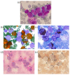
Bone marrow (BM) morphological examination with Romanowsky–Giemsa staining (a) and cytochemical examination for MPO (b), lipids (Sudan black staining) (c), glycogen (PAS reaction) (d) and non-specific esterase (e). Identified cells are marked as follows: B—blast, E—eosinophil, L—lymphocyte, Me—metamyelocyte, Mo—monocyte, My—myelocyte, N—neutrophil, P—promonocyte, 100× magnification.
Figure 2
Figure 2
Immunophenotypic description of the bone marrow (BM). A BM overview is presented in the “gray” panel; a brief description of early myeloid blasts is presented in the “red” panel; two populations of γδ-T lymphocytes are shown in blue and violet, while maturation of monocytic cells is shown in orange.
Figure 3
Figure 3
Lymph node biopsy examination with hematoxylin-eosin staining at 100× (a) and 400× (b) magnification, and immunohistochemical examination at 400× for MPO (c) and CD3 (d).
Figure 4
Figure 4
Conventional G-banded karyotype analysis showing the t(3;11)(q21;q23) translocation in whole BM. Rearranged chromosomes are marked with arrows.
Figure 5
Figure 5
CBL gene, exon 8 Sanger sequencing showing c.1186T>C, p.Cys396Arg missense variant (highlighted in yellow) in the BM (a), lymph node (b), and it’s absence in the nails (c).
Figure 6
Figure 6
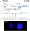
Molecular findings on SET::NUP214 fusion in the BM: structure of SET::NUP214 fusion transcript in RNAseq data as depicted using the Arriba algorithm (a) and Sanger validation (SET::NUP214 exon 6—exon 18 junction is marked) (b); NUP214 gene rearrangement with 5′-partial deletion confirmed using FISH with NUP214 breakapart probe (CytoCell), 63× magnification, wild-type NUP214 is yellow, rearranged NUP214 3’-portion is green (c).
Figure 7
Figure 7
TCR repertoire: fraction of TRB (a) and TRD (b) clonal rearrangements in lymph node. BM—bone marrow aspirate, LN—lymph node biopsy. BM populations are as follows: B—early myeloid blasts (CD45+CD33+CD14), Mo—maturing and mature monocytes (CD45+CD33+CD14+), γδ-T1—immature γδ-T lymphocytes considered leukemic (CD45dimCD3dim), γδ-T2—more mature γδ-T lymphocytes considered normal (CD45highCD3high).
Figure 8
Figure 8
CT scan data. (a) Initial CT scan on admission with massive lymphoproliferative syndrome. Red circles indicate lymph node conglomerates. (b) Computed tomography at 180 days after hematopoietic stem cell transplantation; no lymphadenopathy and recession of the lymphoproliferative syndrome are seen.
Figure 9
Figure 9
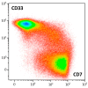
Continuity of the flow cytometric pattern of myeloid and T-lineage cells in BM of described patient, density plot of CD7 and CD33 expression (represented by color scale from red to blue). Mature neutrophils, erythroid precursors, NK cells, and B lymphocytes are excluded.

References

    1. Siegel R.L., Miller K.D., Wagle N.S., Jemal A. Cancer statistics, 2023. CA Cancer J. Clin. 2023;73:17–48. doi: 10.3322/caac.21763. - DOI - PubMed
    1. Noone A.M., Howlader N., Krapcho M., Miller D., Brest A., Yu M., Ruhl J., Tatalovich Z., Mariotto A., Lewis D.R., et al. SEER Cancer Statistics Review, 1975–2015. National Cancer Institute; Bethesda, MD, USA: 2018. [(accessed on 10 January 2023)]. Available online: https://seer.cancer.gov/csr/1975_2015/
    1. Phan A., Veldman R., Lechowicz M.J. T-cell Lymphoma Epidemiology: The Known and Unknown. Curr. Hematol. Malig. Rep. 2016;11:492–503. doi: 10.1007/s11899-016-0353-y. - DOI - PubMed
    1. Vose J., Armitage J., Weisenburger D., International T.C.L.P. International peripheral T-cell and natural killer/T-cell lymphoma study: Pathology findings and clinical outcomes. J. Clin. Oncol. 2008;26:4124–4130. doi: 10.1200/JCO.2008.16.4558. - DOI - PubMed
    1. Hathuc V., Kreisel F. Genetic Landscape of Peripheral T-Cell Lymphoma. Life. 2022;12:410. doi: 10.3390/life12030410. - DOI - PMC - PubMed

Publication types

MeSH terms

Substances