Review
. 2023 Sep 26;24(19):14582.
doi: 10.3390/ijms241914582.

The Involvement of Neuroinflammation in the Onset and Progression of Parkinson's Disease

Affiliations
Review

The Involvement of Neuroinflammation in the Onset and Progression of Parkinson's Disease

Anamaria Jurcau et al. Int J Mol Sci. .

Abstract

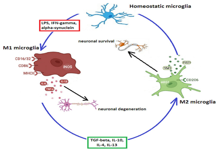
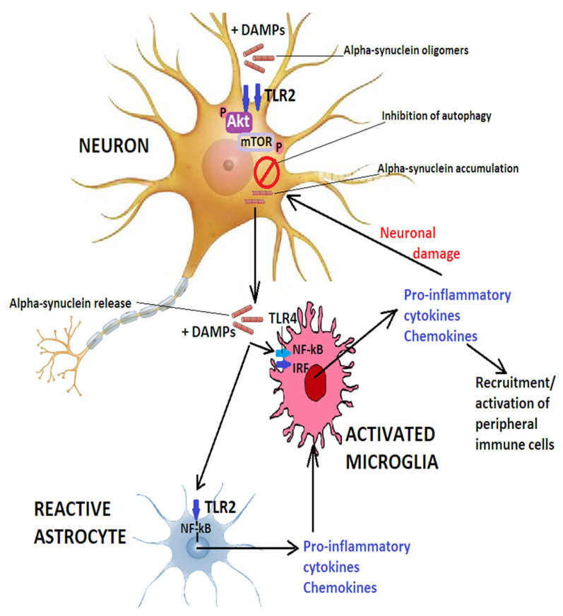
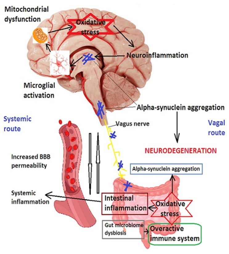
Parkinson's disease is a neurodegenerative disease exhibiting the fastest growth in incidence in recent years. As with most neurodegenerative diseases, the pathophysiology is incompletely elucidated, but compelling evidence implicates inflammation, both in the central nervous system and in the periphery, in the initiation and progression of the disease, although it is not yet clear what triggers this inflammatory response and where it begins. Gut dysbiosis seems to be a likely candidate for the initiation of the systemic inflammation. The therapies in current use provide only symptomatic relief, but do not interfere with the disease progression. Nonetheless, animal models have shown promising results with therapies that target various vicious neuroinflammatory cascades. Translating these therapeutic strategies into clinical trials is still in its infancy, and a series of issues, such as the exact timing, identifying biomarkers able to identify Parkinson's disease in early and pre-symptomatic stages, or the proper indications of genetic testing in the population at large, will need to be settled in future guidelines.

Keywords: M1 phenotype; M2 phenotype; Parkinson’s disease; astroglia; gut dysbiosis; microglia; neuroinflammation; signaling pathways; therapy; α-synuclein.


Conflict of interest statement

The authors declare no conflict of interest.

Figures

Figure 1
Figure 1
M1 and M2 microglial phenotypes. Normally, microglia exhibit the resting, homeostatic phenotype. Pathogens, such as LPS, α-synuclein, or other misfolded proteins, as well as interferon-γ, shift microglia toward the M1 pro-inflammatory phenotype, which produces ROS and pro-inflammatory cytokines such as IL-6, IL-1β, NOS, or TNF-α, with subsequent neuronal degeneration. Cytokines such as TGF-β, IL-10, IL-4, or IL-13 lead to the transition of the microglial M1 phenotype to the anti-inflammatory M2 phenotype, which promotes neuronal survival by secreting anti-inflammatory cytokines such as FIZZ1 or YM1. Abbreviations: Arg1—arginase-1; CD—mannose receptors (cluster of differentiation); FIZZ1—found in inflammatory zone protein 1; IFN—interferon; IL—interleukin; LPS—lipopolysaccharides; MHC—major histocompatibility complex molecules; NOS—nitric oxide synthase; TGF—tumor growth factor; TNF—tumor necrosis factor; Ym1—chitinase-3 like-3 (in rodents known as YM1).
Figure 2
Figure 2
The role of TLR2 and TLR4 in PD. Oligomeric α-synuclein or other DAMPs activate neuronal TLR2 and inhibit autophagy via the Akt/mTOR pathway. This inhibits α-synuclein clearance and causes the release of the misfolded protein which, together with other DAMPs, activate microglia via TLR2 and TLR4 and leads to translocation of IRF and NF-κB to the nucleus and secretion of pro-inflammatory cytokines and chemokines that exacerbate neuronal damage and may additionally recruit peripheral immune cells to the CNS. A-synuclein released by neurons, together with other DAMPs, also triggers astrocyte activation via TLR2 and induces the production of pro-inflammatory mediators which can further contribute to microglial activation. Abbreviations: DAMPs—damage-associated molecular patterns; IRF—interferon regulatory factor; NF-κB—nuclear factor kappa light-chain-enhancer of activated B cells; TLR—toll-like receptor.
Figure 3
Figure 3
The gut–brain axis in Parkinson’s disease. Alterations in the gut microbiome lead to activation of the immune system and increase in oxidative stress, resulting in enhanced permeability of the intestinal epithelium, which allows bacterial products and α-synuclein to spread via systemic circulation and/or the vagal route from the enteric plexuses to the CNS, where they induce microglial activation, mitochondrial dysfunction, promote oxidative stress and neuroinflammation, potentiating α-synuclein aggregation, and resulting in neurodegeneration. BBB—blood–brain barrier.

References

    1. Dauer W., Przedborski S. Parkinson’s disease: Mechanisms and models. Neuron. 2003;39:889–909. doi: 10.1016/S0896-6273(03)00568-3. - DOI - PubMed
    1. Dorsey E.R., Sherer T., Okun M.S., Bloem B.R. The emerging evidence of the Parkinson pandemic. J. Park. Dis. 2018;8:S3–S8. doi: 10.3233/JPD-181474. - DOI - PMC - PubMed
    1. GBD 2015 Neurological Disorders Collaborator Group Global, regional, and national burden of neurological disorders during 1990–2015: A systematic analysis for the Global Burden of Disease Study. Lancet Neurol. 2017;16:877–897. doi: 10.1016/S1474-4422(17)30299-5. - DOI - PMC - PubMed
    1. Dorsey E., Bloem B.R. The parkinson pandemic—A call to action. JAMA Neurol. 2018;75:9–10. doi: 10.1001/jamaneurol.2017.3299. - DOI - PubMed
    1. Jankovic J. Parkinson’s disease: Clinical features and diagnosis. J. Neurol. Neurosurg. Psychiatry. 2008;79:368–376. doi: 10.1136/jnnp.2007.131045. - DOI - PubMed