. 2023 Sep 27;24(19):14620.
doi: 10.3390/ijms241914620.

Synergistic Therapeutic Potential of Dual 3D Mesenchymal Stem Cell Therapy in an Ischemic Hind Limb Mouse Model

Affiliations

Synergistic Therapeutic Potential of Dual 3D Mesenchymal Stem Cell Therapy in an Ischemic Hind Limb Mouse Model

Dong-Sik Chae et al. Int J Mol Sci. .

Abstract

Three-dimensional (3D) culture systems have been widely used to promote the viability and metabolic activity of mesenchymal stem cells (MSCs). The aim of this study was to explore the synergistic benefits of using dual 3D MSC culture systems to promote vascular regeneration and enhance therapeutic potential. We used various experimental assays, including dual 3D cultures of human adipose MSCs (hASCs), quantitative reverse transcription polymerase chain reaction (qRT-PCR), in vitro cell migration, Matrigel tube network formation, Matrigel plug assay, therapeutic assays using an ischemic hind limb mouse model, and immunohistochemical analysis. Our qRT-PCR results revealed that fibroblast growth factor 2 (FGF-2), granulocyte chemotactic protein-2 (GCP-2), and vascular endothelial growth factor-A (VEGF-A) were highly upregulated in conventional 3D-cultured hASCs (ASC-3D) than in two-dimensional (2D)-cultured hASCs. Hepatocyte growth factor (HGF), insulin-like growth factor-1 (IGF-1), and stromal-cell-derived factor-1 (SDF-1) showed higher expression levels in cytokine-cocktail-based, 3D-cultured hASCs (ASC-3Dc). A conditioned medium (CM) mixture of dual 3D ASCs (D-3D; ASC-3D + ASC-3Dc) resulted in higher migration and Matrigel tube formation than the CM of single 3D ASCs (S-3D; ASC-3D). Matrigel plugs containing D-3D contained more red blood cells than those containing S-3D. D-3D transplantation into ischemic mouse hind limbs prevented limb loss and augmented blood perfusion when compared to S-3D transplantation. Transplanted D-3D also revealed a high capillary density and angiogenic cytokine levels and transdifferentiated into endothelial-like cells in the hind limb muscle. These findings highlight the benefits of using the dual 3D culture system to optimize stem-cell-based therapeutic strategies, thereby advancing the therapeutic strategy for ischemic vascular disease and tissue regeneration.

Keywords: 3D culture systems; adipose mesenchymal stem cells; angiogenic potential; regenerative medicine; tissue engineering.


Conflict of interest statement

The authors declare no conflict of interest.

Figures
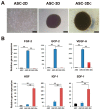
Figure 1
Figure 1
Analysis of morphology and proangiogenic gene expression in two-dimensional (2D) or three-dimensional (3D) adipose-derived mesenchymal stem cells (ASC). (A) Morphology of 2D ASCs, ASC-3D, and 3D-cultured hASCs (ASC-3Dc). Bars = 100 μm. (B) Quantitative reverse transcription polymerase chain reaction (qRT-PCR) was used to measure gene expression patterns of multiple factors. Individual values were normalized to the expression level of glyceraldehyde-3-phosphate dehydrogenase (GAPDH). ** p < 0.01; n = 4 per group.
Figure 2
Figure 2
In vitro and in vivo angiogenic properties of dual 3D ASCs (D-3D). (A) Representative photograph of scratch wound migration. Bars = 100 μm. (B) The culture medium (CM) of D-3D highly improved wound closure through human dermal fibroblasts (HDFs) when compared to the CM of single 3D ASCs (S-3D). * p < 0.05, ** p < 0.01; n = 5 per group. (C) Representative photograph of Matrigel tube formation. Bars = 100 μm. (D) Quantification of branching point and tube length. The CM of D-3D significantly increased the formation of a tubular structure when compared to the CM of S-3D. ** p < 0.01; n = 5 per group. (E) Representative photograph of Matrigel plugs injected with S-3D and D-3D at 14 days after cell injection. (F) Quantification of hemoglobin content. D-3D injection significantly increased the hemoglobin content when compared to S-3D. ** p < 0.01; n = 4 per group.
Figure 3
Figure 3
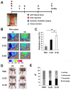
Analysis of the angiogenic therapeutic effects of D-3D in an ischemic hind limb model. (A) A schematic of the experimental protocol for ischemic hind limb surgery, cell transplantation, LDPI analysis, and subsequent tissue collection. (B) Representative LDPI images depicting the measurement of blood flow recovery in ischemic hind limbs following cell injection. (C) Quantitative analysis of blood perfusion conducted 2 weeks after cell injection. n = 7 per group. ** p < 0.01; * p < 0.05. (D) Representative images of limb salvage, limb necrosis, or limb loss after cell transplantation. (E) Quantitative analysis of limb salvage, limb necrosis, or limb loss after cell injection.
Figure 4
Figure 4
Analysis of the therapeutic mechanism. (A) Representative images showing the capillary density in hind limb tissues at 4 weeks after cell injection. The blue stain represents nuclear 4′,6-diamidino-2-phenylindole (DAPI), while the green stain represents interleukin beta 4 (ILB4). Bars = 500 μm. (B) Quantitative analysis of the capillary density in hind limb tissues after cell injection. Statistical analysis showed significant differences between groups, indicated as ** p < 0.01 and * p < 0.05. n = 6 per group. (C) Histological images of hind limb tissues stained with hemolysin and eosin (H&E) at 15 days after cell injection. Bars = 200 μm. (D) Analysis of angiogenic factor expression in hind limb tissues 3 days after cell transplantation. n = 7 per group. ** p < 0.01; * p < 0.05.
Figure 5
Figure 5
Engraftment and endothelial differentiation property of D-3D in an ischemic hind limb model. Representative images of co-localized Dil-labeled D-3D in the ischemic hind limb 4 weeks after cell injection. Nuclei were stained with DAPI (blue). ILB4 (green), Dil (red). Arrows indicate ILB4 and Dil double-positive cells, suggesting endothelial differentiation of D-3D. Bars = 500 μm.
Figure 6
Figure 6
D-3D-based therapy strategy to promote angiogenesis. D-3D therapy exhibited superior angiogenic activity in vitro when compared to S-3D therapy. In a mouse HLI model, D-3D therapy resulted in more substantial limb ischemia repair compared to S-3D therapy. Mechanistically, the injected D-3D therapy induced higher neovascularization. Arrows indicating an increase.

Similar articles

Cited by

References

    1. Simon F., Oberhuber A., Floros N., Busch A., Wagenhauser M.U., Schelzig H., Duran M. Acute Limb Ischemia-Much More than Just a Lack of Oxygen. Int. J. Mol. Sci. 2018;19:374. doi: 10.3390/ijms19020374. - DOI - PMC - PubMed
    1. Varu V.N., Hogg M.E., Kibbe M.R. Critical limb ischemia. J. Vasc. Surg. 2010;51:230–241. doi: 10.1016/j.jvs.2009.08.073. - DOI - PubMed
    1. Atluri P., Woo Y.J. Pro-angiogenic cytokines as cardiovascular therapeutics: Assessing the potential. BioDrugs Clin. Immunother. Biopharm. Gene Ther. 2008;22:209–222. doi: 10.2165/00063030-200822040-00001. - DOI - PubMed
    1. Henry T.D., Annex B.H., McKendall G.R., Azrin M.A., Lopez J.J., Giordano F.J., Shah P.K., Willerson J.T., Benza R.L., Berman D.S., et al. The VIVA trial: Vascular endothelial growth factor in Ischemia for Vascular Angiogenesis. Circulation. 2003;107:1359–1365. doi: 10.1161/01.CIR.0000061911.47710.8A. - DOI - PubMed
    1. Borlongan C.V. Age of PISCES: Stem-cell clinical trials in stroke. Lancet. 2016;388:736–738. doi: 10.1016/S0140-6736(16)31259-4. - DOI - PubMed

MeSH terms

Substances

LinkOut - more resources