. 2023 Sep 27;24(19):14629.
doi: 10.3390/ijms241914629.

NET Formation Was Reduced via Exposure to Extremely Low-Frequency Pulsed Electromagnetic Fields

Affiliations

NET Formation Was Reduced via Exposure to Extremely Low-Frequency Pulsed Electromagnetic Fields

Caren Linnemann et al. Int J Mol Sci. .

Abstract

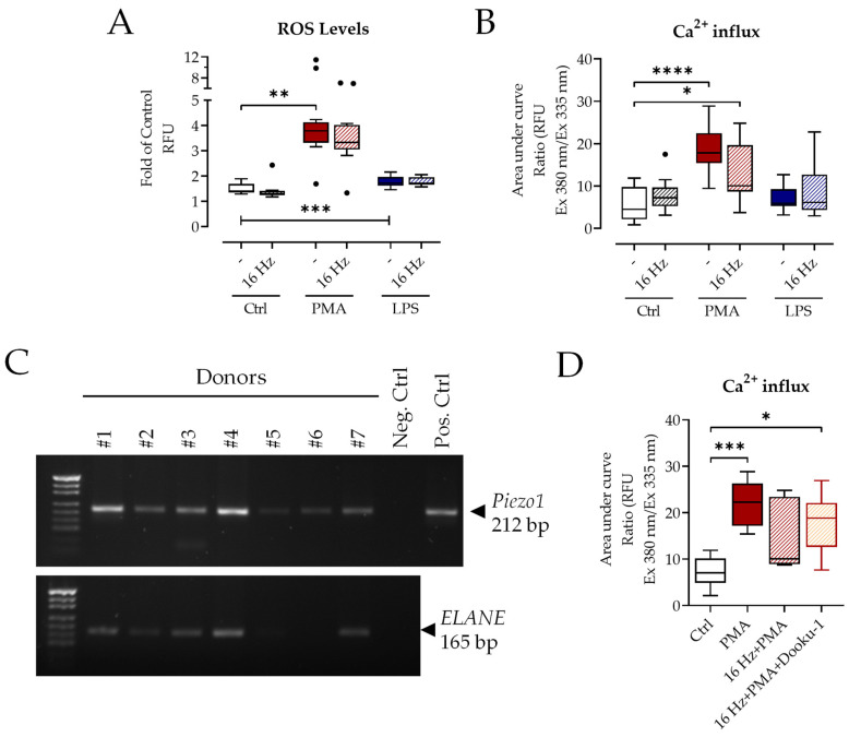
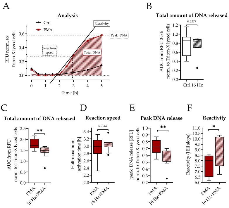
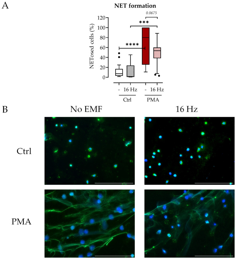
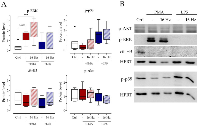
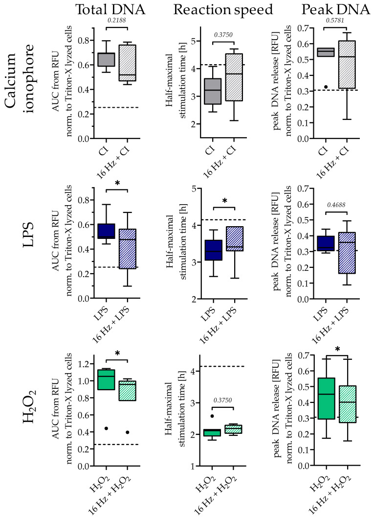
Fracture-healing is a highly complex and timely orchestrated process. Non-healing fractures are still a major clinical problem and treatment remains difficult. A 16 Hz extremely low-frequency pulsed electromagnetic field (ELF-PEMF) was identified as non-invasive adjunct therapy supporting bone-healing by inducing reactive oxygen species (ROS) and Ca2+-influx. However, ROS and Ca2+-influx may stimulate neutrophils, the first cells arriving at the wounded site, to excessively form neutrophil extracellular traps (NETs), which negatively affects the healing process. Thus, this study aimed to evaluate the effect of this 16 Hz ELF-PEMF on NET formation. Neutrophils were isolated from healthy volunteers and exposed to different NET-stimuli and the 16 Hz ELF-PEMF. NETs were quantified using Sytox Green Assay and immunofluorescence, Ca2+-influx and ROS with fluorescence probes. In contrast to mesenchymal cells, ELF-PEMF exposure did not induce ROS and Ca2+-influx in neutrophils. ELF-PEMF exposure did not result in basal or enhanced PMA-induced NET formation but did reduce the amount of DNA released. Similarly, NET formation induced by LPS and H2O2 was reduced through exposure to ELF-PEMF. As ELF-PEMF exposure did not induce NET release or negatively affect neutrophils, the ELF-PEMF exposure can be started immediately after fracture treatment.

Keywords: EMF; fracture healing; neutrophil extracellular traps; neutrophils.


Conflict of interest statement

The authors declare no conflict of interest. Sachtleben GmbH provided the ELF-PEMF devices with the technical support but was not involved in the study design or the data evaluation.

Figures

Figure 1
Figure 1
Exposure to 16 Hz ELF-PEMF does not change ROS formation or Ca2+-influx. Neutrophils were stimulated through ±100 nM PMA or ±25 µg/mL LPS after ±7 min exposure to 16 Hz ELF-PEMF. (A) Amount of total ROS formed was determined via DCFH-DA assay, N = 5, n = 3. (B) Ca2+-influx was quantified using Fura-2-AM measurement, N = 5, n = 3. (C) Results of Piezo1 and ELANE PCR for neutrophils from seven different donors. (D) Ca2+-influx was quantified via Fura-2-AM measurement ± Piezo1 inhibitor Dooku-1 (20 µM), N = 5, n = 3. * p < 0.05, ** p < 0.01, *** p < 0.001, **** p < 0.0001, as determined via two-way ANOVA (A,B) or non-parametric Kruskal–Wallis test (D). Dots in the box plots indicate outliers. White bars: no additional stimulation, red bars: PMA stimulation, blue bars: LPS stimulation, shading indicates exposure to 16 Hz ELF-PEMF, orange indicates addition of Piezo-1 inhibitor.
Figure 2
Figure 2
The 16 Hz EMF does not induce NET formation. (A) Time course of the Sytox Green assay of Ctrl- and PMA-stimulated neutrophils (no ELF-PEMF exposure), different analysis parameters are highlighted in the graph, shading indicates the area under the curve which represents the total amount of released DNA. (B) Total amount of DNA released obtained via the Sytox Green assay, ±7 min exposure of 16 Hz ELF-PEMF. N = 10, n = 3. (CF) Different analysis parameters calculated from the Sytox Green assay with the addition of 100 nM PMA ±7 min of 16 Hz ELF-PEMF. N = 10, n = 3. (C) Total amount of released DNA. (D) Reaction speed. (E) Peak DNA release. (F) Reactivity. Statistical analysis was carried out using Wilcoxon’s test with * p < 0.05, ** p < 0.01. Dots in the box plots indicate outliers.
Figure 3
Figure 3
The 16 Hz ELF-PEMF exposure does not induce NET formation. Neutrophils were stimulated ±100 nM PMA after ±7 min exposure to 16 Hz ELF-PEMF. After 240 min, immunofluorescent staining for NETs was performed. (A) The number of NETosed cells was quantified from immunofluorescence images using the ImageJ software (Version 1.53), shading indicates exposure to 16 Hz ELF-PEMF, N = 3, n = 5. (B) Exemplary images of immunofluorescent stainings: Blue: DNA (Hoechst 33342); green: myeloperoxidase. Scale bar: 200 µm. *** p < 0.001 and **** p < 0.0001 as determined via two-way ANOVA. Dots in the box plots indicate outliers.
Figure 4
Figure 4
The 16 Hz ELF-PEMF exposure does not change MAPK activation. Neutrophils were stimulated ±100 nM PMA or ±25 µg/mL LPS after ±7 min exposure to 16 Hz ELF-PEMF, shading indicates exposure to 16 Hz ELF-PEMF. (A) Protein levels of indicated proteins determined via Western blot after 1.5 h of stimulation. (B) Exemplary blot images. N = 4, n = 2. ** p < 0.01, as determined by non-parametric Kruskal–Wallis test. Dots in the box plots indicate outliers.
Figure 5
Figure 5
The 16 Hz ELF-PEMF exposure had a similar effect on NET formation to other stimulants. Total amount of DNA, activation time, and peak DNA release obtained from the Sytox Green assay. Neutrophils were stimulated using ±4 µM CI, ±25 µg/mL LPS, or ±0.003% H2O2 after ±7 min exposure of 16 Hz ELF-PEMF. N = 7, n = 4. Dashed line indicates control group without ELF-PEMF exposure. * p < 0.05, as determined using the Mann–Whitney U test. Dots in the box plots indicate outliers.

Similar articles

Cited by

References

    1. Gomez-Barrena E., Rosset P., Lozano D., Stanovici J., Ermthaller C., Gerbhard F. Bone fracture healing: Cell therapy in delayed unions and nonunions. Bone. 2015;70:93–101. doi: 10.1016/j.bone.2014.07.033. - DOI - PubMed
    1. Karl T., Gussmann A., Storck M. Chronische Wunden—Perspektiven der integrierten Versorgung. Zentralblatt Chir. 2007;132:232–235. doi: 10.1055/s-2007-960751. - DOI - PubMed
    1. Ziegler P., Nussler A.K., Wilbrand B., Falldorf K., Springer F., Fentz A.K., Eschenburg G., Ziegler A., Stöckle U., Maurer E., et al. Pulsed Electromagnetic Field Therapy Improves Osseous Consolidation after High Tibial Osteotomy in Elderly Patients-A Randomized, Placebo-Controlled, Double-Blind Trial. J. Clin. Med. 2019;8:2008. doi: 10.3390/jcm8112008. - DOI - PMC - PubMed
    1. Caliogna L., Medetti M., Bina V., Brancato A.M., Castelli A., Jannelli E., Ivone A., Gastaldi G., Annunziata S., Mosconi M., et al. Pulsed Electromagnetic Fields in Bone Healing: Molecular Pathways and Clinical Applications. Int. J. Mol. Sci. 2021;22:7403. doi: 10.3390/ijms22147403. - DOI - PMC - PubMed
    1. Zhu S., He H., Zhang C., Wang H., Gao C., Yu X., He C. Effects of pulsed electromagnetic fields on postmenopausal osteoporosis. Bioelectromagnetics. 2017;38:406–424. doi: 10.1002/bem.22065. - DOI - PubMed