. 2023 Sep 29;24(19):14749.
doi: 10.3390/ijms241914749.

Altered Extracellular Vesicle miRNA Profile in Prodromal Alzheimer's Disease

Affiliations

Altered Extracellular Vesicle miRNA Profile in Prodromal Alzheimer's Disease

Caterina Visconte et al. Int J Mol Sci. .

Abstract

Extracellular vesicles (EVs) are nanosized vesicles released by almost all body tissues, representing important mediators of cellular communication, and are thus promising candidate biomarkers for neurodegenerative diseases like Alzheimer's disease (AD). The aim of the present study was to isolate total EVs from plasma and characterize their microRNA (miRNA) contents in AD patients. We isolated total EVs from the plasma of all recruited subjects using ExoQuickULTRA exosome precipitation solution (SBI). Subsequently, circulating total EVs were characterized using Nanosight nanoparticle tracking analysis (NTA), transmission electron microscopy (TEM), and Western blotting. A panel of 754 miRNAs was determined with RT-qPCR using TaqMan OpenArray technology in a QuantStudio 12K System (Thermo Fisher Scientific). The results demonstrated that plasma EVs showed widespread deregulation of specific miRNAs (miR-106a-5p, miR-16-5p, miR-17-5p, miR-195-5p, miR-19b-3p, miR-20a-5p, miR-223-3p, miR-25-3p, miR-296-5p, miR-30b-5p, miR-532-3p, miR-92a-3p, and miR-451a), some of which were already known to be associated with neurological pathologies. A further validation analysis also confirmed a significant upregulation of miR-16-5p, miR-25-3p, miR-92a-3p, and miR-451a in prodromal AD patients, suggesting these dysregulated miRNAs are involved in the early progression of AD.

Keywords: Alzheimer’s disease (AD); biomarker; extracellular vesicles; miRNA.


Conflict of interest statement

The authors declare no conflict of interest.

Figures

Figure 1
Figure 1
Characterization of isolated plasma vesicles. (a) Representative trace of nanoparticle tracking analysis (NTA) of total EVs isolated using ExoQuick ULTRA. (b) Plasma, total EVs, and depleted EVs (DEVs) were analyzed by immunoblotting with antibodies against CD81, TGS101, and GM130, as indicated on the right. (c) TEM images revealed the peculiar oval shape of circulating EVs.
Figure 2
Figure 2
MiRNA expression profile in total plasma EVs. (a) Concentration of EV-derived miRNA after using miRNeasy Isolation Kit assessed by Bioanalyzer. (b) Pie chart showing deregulated miRNAs from the total number of miRNAs analyzed. (c) Box plots showing 2−ΔCt. and min and max values for CTRL, non-AD MCI, and AD patients. * p < 0.05; ** p < 0.005.
Figure 3
Figure 3
miRNA expression levels in non-AD MCI, prodromal AD, AD, and CTRL, shown as 2−ΔCt. * p < 0.05; ** p < 0.005.
Figure 4
Figure 4
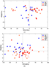
(a) PCA based on the most variant miRNAs in the discovery cohort (p = 0.004). (b) PCA based on the most variant miRNAs in the validation cohort (p = 0.007). The p-value between AD and control samples was computed using a Wilcoxon test applied to the first principal component.
Figure 5
Figure 5
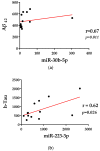
(a) The Spearman correlation analysis between miR-30b-5p and Aβ42. (b) The Spearman correlation analysis between miR-30b-5p and Aβ42.
Figure 6
Figure 6
Network visualization for the enrichment analysis of miR-92a-3p, miR-16-5p, miR-25-3p, and miR-451a in prodromal AD and AD groups. Red/yellow squares represent deregulated miRNAs and pink circles are their target genes, while yellow circles are their lncRNA targets. Big red/yellow circles represent shared interactions.
Figure 7
Figure 7
MiRNA target and pathway prediction. (a) Biological pathway (y-axis) associated with mir-25-3p, mir-92a-3p, mir-16-5p and mir-451a signature. The significance of this association is expressed with –log10 (p-value) and is reported on x-axis. (b) Network visualization for the enrichment analysis of miR-92a-3p, miR-16-5p, miR-25-3p, and miR-451a in prodromal AD and AD group. Pink circles are their targets genes while yellow circles are their lncRNA targets. Big orange circles represent Wnt Signaling pathway; big blue circles show genes involved in phagosomes; red circles show gap-junction-associated genes; green circles represent genes related to synaptic vesicle cycle.

References

    1. Liss J.L., Assunção S.S.M., Cummings J., Atri A., Geldmacher D.S., Candela S.F., Devanand D.P., Fillit H.M., Susman J., Mintzer J., et al. Practical recommendations for timely, accurate diagnosis of symptomatic Alzheimer’s disease (MCI and dementia) in primary care: A review and synthesis. J. Intern. Med. 2021;290:310–334. doi: 10.1111/joim.13244. - DOI - PMC - PubMed
    1. Blennow K. A Review of Fluid Biomarkers for Alzheimer’s Disease: Moving from CSF to Blood. Neurol. Ther. 2017;6:15–24. doi: 10.1007/s40120-017-0073-9. - DOI - PMC - PubMed
    1. Aharon A., Spector P., Ahmad R.S., Horrany N., Sabbach A., Brenner B., Aharon-Peretz J. Extracellular Vesicles of Alzheimer’s Disease Patients as a Biomarker for Disease Progression. Mol. Neurobiol. 2020;57:4156–4169. doi: 10.1007/s12035-020-02013-1. - DOI - PubMed
    1. Aulston B., Liu Q., Mante M., Florio J., Rissman R.A., Yuan S.H. Extracellular Vesicles Isolated from Familial Alzheimer’s Disease Neuronal Cultures Induce Aberrant Tau Phosphorylation in the Wild-Type Mouse Brain. J. Alzheimer’s Dis. JAD. 2019;72:575–585. doi: 10.3233/JAD-190656. - DOI - PMC - PubMed
    1. Duggan M.R., Lu A., Foster T.C., Wimmer M., Parikh V. Exosomes in Age-Related Cognitive Decline: Mechanistic Insights and Improving Outcomes. Front. Aging Neurosci. 2022;14:834775. doi: 10.3389/fnagi.2022.834775. - DOI - PMC - PubMed