Review
. 2023 Oct 4;24(19):14882.
doi: 10.3390/ijms241914882.

The IGF1 Signaling Pathway: From Basic Concepts to Therapeutic Opportunities

Affiliations
Review

The IGF1 Signaling Pathway: From Basic Concepts to Therapeutic Opportunities

Haim Werner. Int J Mol Sci. .

Abstract

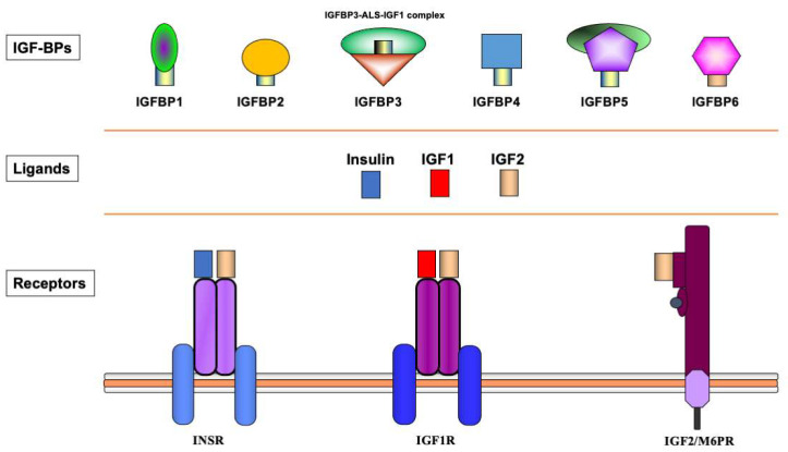
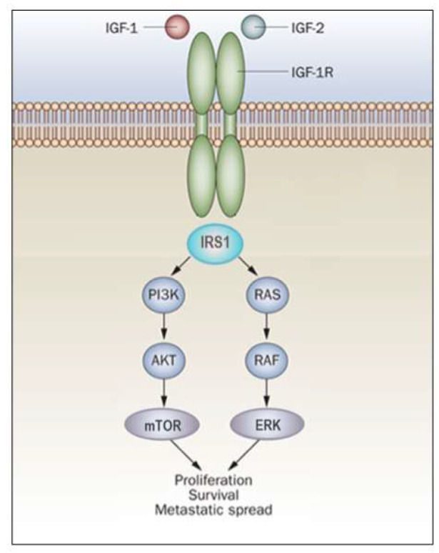
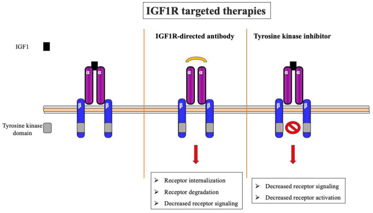
Insulin-like growth factor 1 (IGF1) is a peptide growth factor with important functions in multiple aspects of growth, development and metabolism. The biological actions of IGF1 are mediated by the IGF1 receptor (IGF1R), a cell-surface protein that is evolutionarily related to the insulin receptor (InsR). The effects of IGF1 are moderated by a group of binding proteins (IGFBPs) that bind and transport the ligand in the circulation and extracellular fluids. In mechanistic terms, IGF1R function is linked to the MAPK and PI3K signaling pathways. Furthermore, IGF1R has been shown to migrate to cell nucleus, where it functions as a transcriptional activator. The co-localization of IGF1R and MAPK in the nucleus is of major interest as it suggests novel mechanistic paradigms for the IGF1R-MAPK network. Given its potent anti-apoptotic and pro-survival roles, and in view of its almost universal pattern of expression in most types of cancer, IGF1R has emerged as a promising molecular target in oncology. The present review article provides a concise overview of key scientific developments in the research area of IGF and highlights a number of more recent findings, including its nuclear migration and its interaction with oncogenes and tumor suppressors.

Keywords: IGF1 receptor; MAPK; insulin-like growth factor-1 (IGF1); nuclear translocation; p53.


Conflict of interest statement

The author declare no conflict of interest.

Figures

Figure 1
Figure 1
Schematic representation of the insulin-IGF network. The insulin-IGF family includes three ligands, three receptors and at least six IGFBPs. Experimental analyses provided evidence of cross-reactivity between ligands and receptors. Hence, IGF2 binds with high affinity to the InsR. In addition, both IGF1 and IGF2 activate hybrid receptors, formed by an IGF1R hemireceptor in association with an InsR hemireceptor (not shown). The IGF2/mannose 6-phosphate receptor is responsible for IGF2 recycling and is not involved in signaling. Most IGF1 in the circulation is bound to IGFBP3, which forms a multi-complex with an acid-labile subunit (ALS) and protects IGF1 from proteolytic degradation.
Figure 2
Figure 2
IGF1R signal transduction. The biological actions of IGF1 and IGF2 are transduced by the IGF1R and moderated by a family of at least six IGFBPs. Ligands bind with high affinity to the extracellular portion of IGF1R, and stimulate autophosphorylation of its tyrosine kinase (TK) domain. Upon activation of the IGF1R, IRSs become phosphorylated, with ensuing activation of two cascades, the RAS-MAP kinase (or ERK) and the PI3K-PDK1-Akt/PKB networks. The net consequence of the activation of these pathways is a boost in proliferation and a decrease in apoptosis.
Figure 3
Figure 3
IGF1R-targeted therapies. Schematic representation of two of the most common approaches for IGF1R targeting: anti-IGF1R monoclonal antibodies (center) and small-molecular-weight IGF1R tyrosine kinase inhibitors (right). Blocking of IGF1R by specific antibodies (usually against the extracellular domain) leads to a decrease in ligand binding and IGF1R activation with ensuing receptor internalization and degradation. Tyrosine kinase inhibitors abrogate IGF1R activation and signaling, without major effects on IGF1R expression.

References

    1. Salmon W.D., Daughaday W.H. A hormonally controlled serum factor which stimulates sulfate incorporation by cartilage in vitro. J. Lab. Clin. Med. 1957;49:825–836. - PubMed
    1. Salmon W.D.J., DuVall M.R. In vitro stimulation of leucine incorporation into muscle and cartilage protein by a serum fraction with sulfation factor activity: Differentiation of effects from those of growth hormone and insulin. Endocrinology. 1970;87:1168–1180. doi: 10.1210/endo-87-6-1168. - DOI - PubMed
    1. Daughaday W.H., Hall K., Raben M.S., Salmon W.D.J., van den Brande J.L., van Wyk J.J. Somatomedin: Proposed designation for sulphation factor. Nature. 1972;235:107. doi: 10.1038/235107a0. - DOI - PubMed
    1. LeRoith D., Bondy C., Yakar S., Liu J.-L., Butler A. The Somatomedin hypothesis: 2001. Endocrine Rev. 2001;22:53–74. doi: 10.1210/edrv.22.1.0419. - DOI - PubMed
    1. Dulak N.C., Temin H.M. Multiplication-stimulating activity for chicken embryo fibroblasts from rat liver cell conditioned medium: A family of small polypeptides. J. Cell Physiol. 1973;81:161–170. doi: 10.1002/jcp.1040810205. - DOI - PubMed

MeSH terms

Substances