. 2023 Oct 6;24(19):14962.
doi: 10.3390/ijms241914962.

Neonatal Exposure to Lipopolysaccharide Promotes Neurogenesis of Subventricular Zone Progenitors in the Developing Neocortex of Ferrets

Affiliations

Neonatal Exposure to Lipopolysaccharide Promotes Neurogenesis of Subventricular Zone Progenitors in the Developing Neocortex of Ferrets

Kazuhiko Sawada et al. Int J Mol Sci. .

Abstract

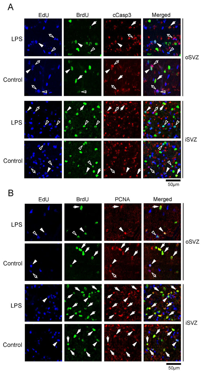
Lipopolysaccharide (LPS) is a natural agonist of toll-like receptor 4 that serves a role in innate immunity. The current study evaluated the LPS-mediated regulation of neurogenesis in the subventricular zone (SVZ) progenitors, that is, the basal radial glia and intermediate progenitors (IPs), in ferrets. Ferret pups were subcutaneously injected with LPS (500 μg/g of body weight) on postnatal days (PDs) 6 and 7. Furthermore, 5-ethynyl-2'-deoxyuridine (EdU) and 5-bromo-2'-deoxyuridine (BrdU) were administered on PDs 5 and 7, respectively, to label the post-proliferative and proliferating cells in the inner SVZ (iSVZ) and outer SVZ (oSVZ). A significantly higher density of BrdU single-labeled proliferating cells was observed in the iSVZ of LPS-exposed ferrets than in controls but not in post-proliferative EdU single-labeled and EdU/BrdU double-labeled self-renewing cells. BrdU single-labeled cells exhibited a lower proportion of Tbr2 immunostaining in LPS-exposed ferrets (22.2%) than in controls (42.6%) and a higher proportion of Ctip2 immunostaining in LPS-exposed ferrets (22.2%) than in controls (8.6%). The present findings revealed that LPS modified the neurogenesis of SVZ progenitors. Neonatal LPS exposure facilitates the proliferation of SVZ progenitors, followed by the differentiation of Tbr2-expressing IPs into Ctip2-expressing immature neurons.

Keywords: Ctip2; Tbr2; carnivores; intermediate progenitors; toll-like receptor.


Conflict of interest statement

There are no conflicts of interest.

Figures

Figure 1
Figure 1
Gross images of the brains of LPS-exposed and control ferrets on postnatal day 7. (A) Dorsal view of the brain. (B) Lateral view of the left side of the brain. (C) Coronal hematoxylin-stained sections are shown from left to right in the rostrocaudal order. White asterisk indicates the inner subventricular zone (iSVZ). Black asterisk indicates the outer subventricular zone (oSVZ). Amg, amygdaloid complex; cc, corpus callosum; Cb, cerebellum; Cd, caudate nucleus; cns, coronal sulcus; CP, cortical plate; gcc, genu of corpus callosum; Hip, hippocampus; Hyp, Hypothalamus; ic, internal capsule; Ln, lentiform nucleus; ls, lateral sulcus; rf, rhinal fissure; rs, rhinal sulcus; rsss, rostral suprasylvian sulcus; Th, thalamus.
Figure 2
Figure 2
EdU and BrdU labeling in the premature cortex in the LPS-exposed and control ferrets on postnatal day 7. (A) Low-magnification images of the outer subventricular zone (oSVZ) and inner subventricular zone (iSVZ). Positions for capturing immunofluorescent images are shown in Figure S1A. (B) High-magnification images of the oSVZ and iSVZ. Positions for capturing immunofluorescent images are shown in Figure S1B. Open arrowheads indicate EdU/BrdU double-labeled cells. VZ, ventricular zone.
Figure 3
Figure 3
Densities of EdU single-, BrdU single-, and EdU/BrdU double-labeled cells in the premature cortex of the LPS-exposed and control ferrets on postnatal day 7. (A) Outer subventricular zone (oSVZ). (B) Inner subventricular zone (iSVZ). Data are shown as means ± standard deviations. Significance is indicated using Scheffe’s test at * p < 0.001; number of ferrets = 3 per group.
Figure 4
Figure 4
Immunofluorescence for cleaved caspase 3 (cCasp3) and proliferating cell nuclear antigen (PCNA) with EdU and BrdU labeling in the premature cortex of the LPS-exposed and control ferrets on postnatal day 7. Positions for capturing immunofluorescent images are shown in Figure S1B. (A) cCasp3 immunofluorescence with EdU and BrdU labeling in the outer and inner subventricular zones (oSVZ and iSVZ). Open arrowheads, EdU/BrdU double-labeled cells; closed arrowheads, cCasp3-immunopositive cells with EdU/BrdU double-labeling; open arrows, cCasp3-immunopositive cells with EdU single-labeling; closed arrows, cCasp3-immunopositive cells with BrdU single-labeling. (B) PCNA immunofluorescence with EdU and BrdU labeling in the oSVZ and iSVZ. Open arrowheads, EdU/BrdU double-labeled cells; closed arrowheads, PCNA-immunopositive cells with EdU/BrdU double-labeling; open arrows, PCNA-immunopositive cells with EdU single-labeling; closed arrows, PCNA-immunopositive cells with BrdU single-labeling.
Figure 5
Figure 5
Immunofluorescence for cell cycle markers Ki67 and phosphohistone H3 (PH3) with EdU and BrdU labeling in the premature cortex of the LPS-exposed and control ferrets on postnatal day 7. Positions for capturing immunofluorescent images are shown in Figure S1B. (A) Ki67 immunofluorescence with EdU and BrdU labeling in the outer and inner subventricular zones (oSVZ and iSVZ). Open arrowheads, EdU/BrdU double-labeled cells; closed arrowheads, Ki67-immunopositive cells with EdU/BrdU double-labeling; open arrows, Ki67-immunopositive cells with EdU single-labeling; closed arrows, Ki67-immunopositive cells with BrdU single-labeling. (B) PH3 immunofluorescence with EdU and BrdU labeling in the oSVZ and iSVZ. Open arrowheads, EdU/BrdU double-labeled cells.
Figure 6
Figure 6
Immunofluorescence for Pax6 with EdU and BrdU labeling in the premature cortex of the LPS-exposed and control ferrets on postnatal day 7. Positions for capturing immunofluorescent images are shown in Figure S1B. Closed arrowheads, Pax6-immunopositive cells with EdU/BrdU double-labeling; closed arrows, Pax6-immunopositive cells with BrdU single-labeling. iSVZ, inner subventricular zone; oSVZ, outer subventricular zone.
Figure 7
Figure 7
Immunofluorescence for Tbr2 and Olig2 with EdU and BrdU labeling in the premature cortex of the LPS-exposed and control ferrets on postnatal day 7. Positions for capturing immunofluorescent images are shown in Figure S1B. (A) Tbr2 immunofluorescence with EdU and BrdU labeling in the outer and inner subventricular zones (oSVZ and iSVZ). Open arrowheads, EdU/BrdU double-labeled cells; closed arrowheads, Tbr2-immunopositive cells with EdU/BrdU double-labeling; open arrows, Tbr2-immunopositive cells with EdU single-labeling; closed arrows, Tbr2-immunopositive cells with BrdU single-labeling. (B) Olig2 immunofluorescence with EdU and BrdU labeling in the oSVZ and iSVZ. Open arrowheads, EdU/BrdU double-labeled cells; closed arrowheads, Olig2-immunopositive cells with EdU/BrdU double-labeling; open arrows, Olig2-immunopositive cells with EdU single-labeling; closed arrows, Olig2-immunopositive cells with BrdU single-labeling.
Figure 8
Figure 8
Immunofluorescence for Cux1 and Ctip2 with EdU and BrdU labeling in the premature cortex of the LPS-exposed and control ferrets on postnatal day 7. Positions for capturing immunofluorescent images are shown in Figure S1B. (A) Cux1 immunofluorescence with EdU and BrdU labeling in the outer and inner subventricular zones (oSVZ and iSVZ). Open arrowheads, EdU/BrdU double-labeled cells; closed arrows, Cux1-immunopositive cells with BrdU single-labeling. (B) Ctip2 immunofluorescence with EdU and BrdU labeling in the oSVZ and iSVZ. Open arrowheads, EdU/BrdU double-labeled cells; open arrows, Ctip2-immunopositive cells with EdU single-labeling; closed arrows, Ctip2-immunopositive cells with BrdU single-labeling.
Figure 9
Figure 9
Bar graphs of densities of cells immunostained for various marker antigens in the premature cortex of the LPS-exposed and control ferrets on postnatal day 7. (A) Outer subventricular zone (oSVZ). (B) Inner subventricular zone (iSVZ). cCasp3, cleaved caspase 3; PH3, phosphohistone H3. Data are shown as means ± standard deviations. Significance is indicated using Scheffe’s test at * p < 0.001; number of ferrets = 3 per group.

Similar articles

Cited by

References

    1. Knuesel I., Chicha L., Britschgi M., Schobel S.A., Bodmer M., Hellings J.A., Toovey S., Prinssen E.P. Maternal immune activation and abnormal brain development across CNS disorders. Nat. Rev. Neurol. 2014;10:643–660. doi: 10.1038/nrneurol.2014.187. - DOI - PubMed
    1. Kwon H.K., Choi G.B., Huh J.R. Maternal inflammation and its ramifications on fetal neurodevelopment. Trends Immunol. 2022;43:230–244. doi: 10.1016/j.it.2022.01.007. - DOI - PMC - PubMed
    1. Shook L.L., Sullivan E.L., Lo J.O., Perlis R.H., Edlow A.G. COVID-19 in pregnancy: Implications for fetal brain development. Trends Mol. Med. 2022;28:319–330. doi: 10.1016/j.molmed.2022.02.004. - DOI - PMC - PubMed
    1. Okun E., Griffioen K.J., Lathia J.D., Tang S.C., Mattson M.P., Arumugam T.V. Toll-like receptors in neurodegeneration. Brain Res. Rev. 2009;59:278–292. doi: 10.1016/j.brainresrev.2008.09.001. - DOI - PMC - PubMed
    1. Barak B., Feldman N., Okun E. Toll-like receptors as developmental tools that regulate neurogenesis during development: An update. Front. Neurosci. 2014;8:272. doi: 10.3389/fnins.2014.00272. - DOI - PMC - PubMed

LinkOut - more resources