. 2023 Oct 8;24(19):14996.
doi: 10.3390/ijms241914996.

Frontotemporal Dementia P301L Mutation Potentiates but Is Not Sufficient to Cause the Formation of Cytotoxic Fibrils of Tau

Affiliations

Frontotemporal Dementia P301L Mutation Potentiates but Is Not Sufficient to Cause the Formation of Cytotoxic Fibrils of Tau

Kuang-Wei Wang et al. Int J Mol Sci. .

Abstract

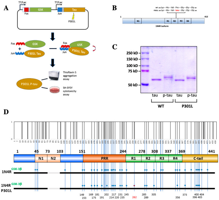
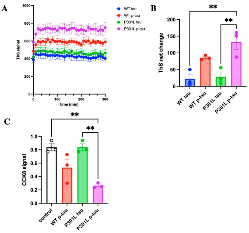
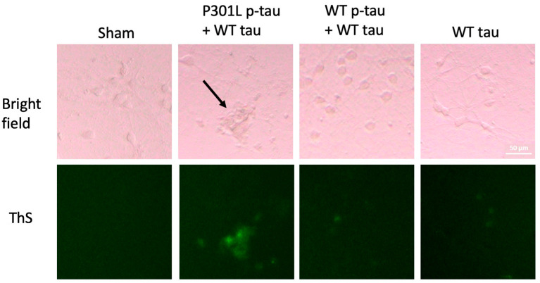
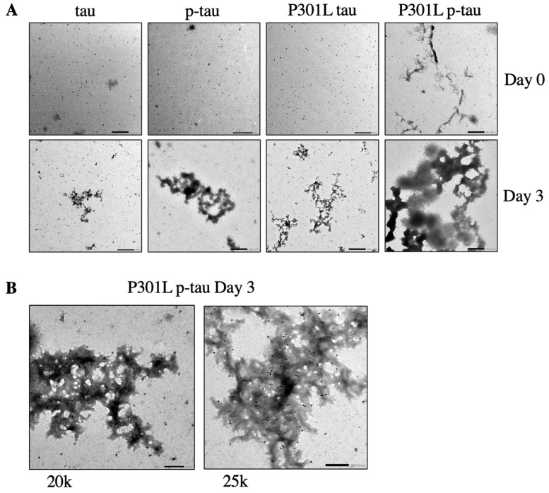
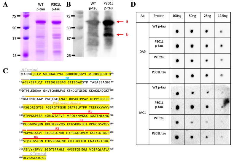
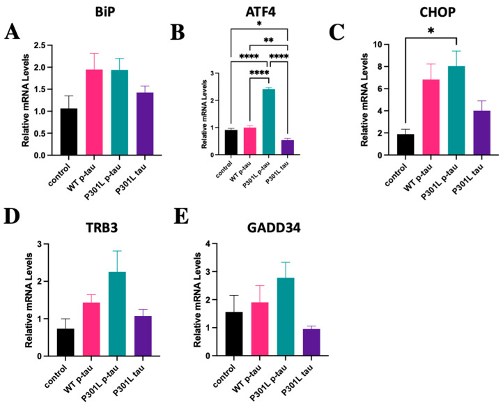
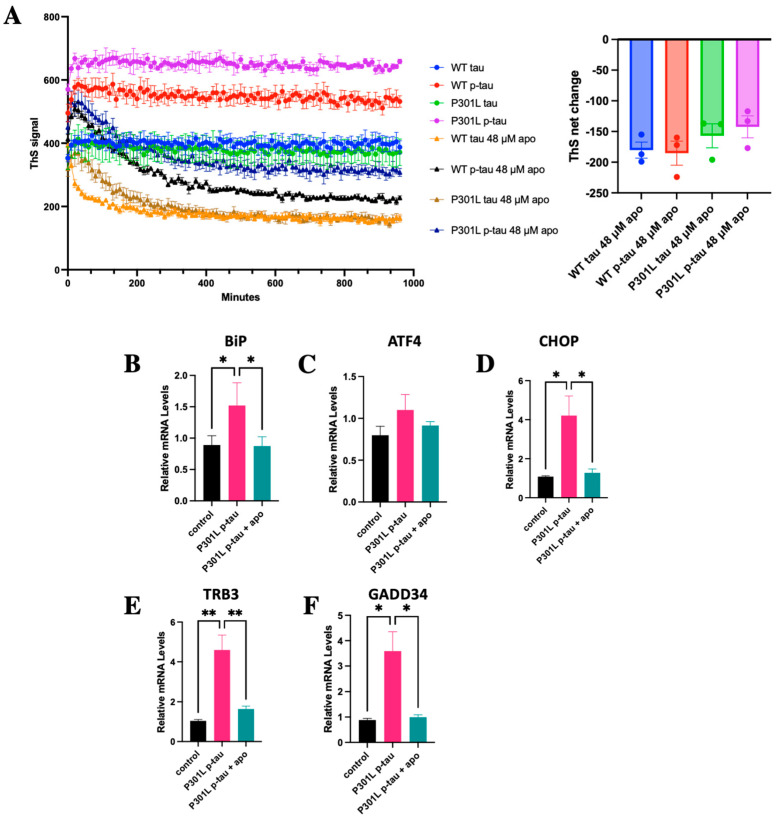
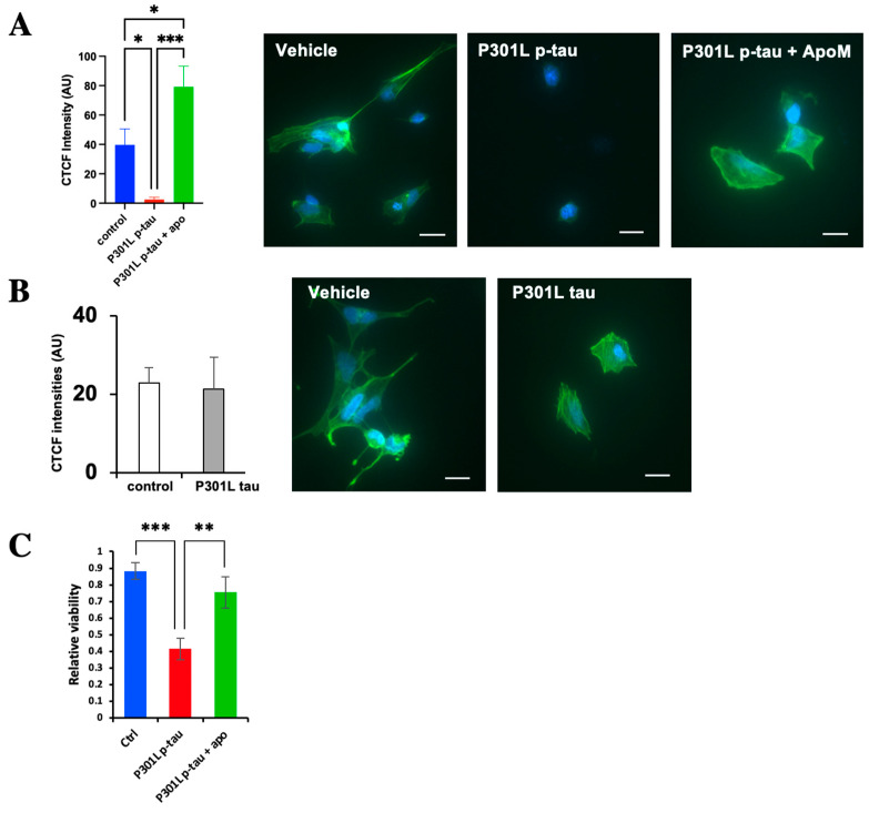
The P301L mutation in tau protein is a prevalent pathogenic mutation associated with neurodegenerative frontotemporal dementia, FTD. The mechanism by which P301L triggers or facilitates neurodegeneration at the molecular level remains unclear. In this work, we examined the effect of the P301L mutation on the biochemical and biological characteristics of pathologically relevant hyperphosphorylated tau. Hyperphosphorylated P301L tau forms cytotoxic aggregates more efficiently than hyperphosphorylated wildtype tau or unphosphorylated P301L tau in vitro. Mechanistic studies establish that hyperphosphorylated P301L tau exacerbates endoplasmic reticulum (ER) stress-associated gene upregulation in a neuroblastoma cell line when compared to wildtype hyperphosphorylated tau treatment. Furthermore, the microtubule cytoskeleton is severely disrupted following hyperphosphorylated P301L tau treatment. A hyperphosphorylated tau aggregation inhibitor, apomorphine, also inhibits the harmful effects caused by P301L hyperphosphorylated tau. In short, the P301L single mutation within the core repeat domain of tau renders the underlying hyperphosphorylated tau more potent in eliciting ER stress and cytoskeleton damage. However, the P301L mutation alone, without hyperphosphorylation, is not sufficient to cause these phenotypes. Understanding the conditions and mechanisms whereby selective mutations aggravate the pathogenic activities of tau can provide pivotal clues on novel strategies for drug development for frontotemporal dementia and other related neurodegenerative tauopathies, including Alzheimer's disease.

Keywords: Alzheimer’s disease; P301L; apomorphine; frontotemporal dementia; hyperphosphorylated tau; tauopathy.


Conflict of interest statement

The authors declare no conflict of interest.

Figures

Figure 1
Figure 1
Production of hyperphosphorylated tau (p-tau). (A) PIMAX plasmid was designed with Fos-GSK and Jun-P301L 1N4R tau and expressed in E. coli. Leucine zipper proteins Fos and Jun modulate the protein interaction between GSK and P301L tau to efficiently produce p-tau. Purified tau and p-tau were assayed with Thioflavin S for aggregation kinetics and administered to SH-SY5Y cells to assay cytotoxic effects. (B) The P301L mutation (at the R2 domain) is known to cause frontotemporal dementia. The P301L missense mutation was introduced into the PIMAX plasmid and confirmed with sequencing. (C) WT tau, WT p-tau, P301L tau, and P301L p-tau purified from E. coli and visualized with Coomassie blue staining. (D) Mass spectrometry analysis of 1N4R tau and 1N4R P301L tau. Dots represent phosphorylated residues. Note that S262 is found to be phosphorylated only in the P301L mutant, whereas several other sites (S46, T50, T111, Y197, S199, T205, T212, S320, S324, S352, T361, T386, S409, and S422) are specific to the wildtype species. Vertical bars on the top represent the S/T/Y phosphorylation sites in the tau sequence.
Figure 2
Figure 2
ThS aggregation assay and SH-SY5Y cell cytotoxicity assay of different tau proteins. (A) Kinetics curves from ThS fluorescence over 5 h. P301L p-tau presents overall the highest ThS signal. (B) Net change of ThS fluorescence was calculated for each sample. P301L p-tau showed the most increased ThS signal. (C) The viability of SH-SY5Y cells treated with 2 μM of tau proteins for 20 h was measured by the CCK8 assay. Quantification with CCK8 shows P301L p-tau is more toxic to cells than WT p-tau and unphosphorylated P301L tau. Error bars obtained from standard error. One-way ANOVA. ** p ≤ 0.01.
Figure 3
Figure 3
P301L p-tau shows strong seeding activity in primary neurons. Mouse primary neurons were treated with a mixture of 0.1 µM p-tau and 0.9 µM tau or 1 µM WT tau alone for three days and then stained by ThS for amyloid structures. P301L p-tau induced the strongest ThS signal and also abnormal cell morphology (black arrow), while WT p-tau had minor effects on tau aggregation and cell shape. Representative images from three technical repeats were shown. Magnification is 20×. Scale bar = 50 µm.
Figure 4
Figure 4
Transmission electron microscopy images of WT p-tau and P301L p-tau aggregation development. (A) PIMAX purified tau, p-tau, P301L tau, P301L p-tau, incubated for 0 days vs. 3 days at 10k magnification. P301L p-tau showed a more organized structure at day 0. Scale bar = 500 nm. (B) Anti-tau immunogold staining of PIMAX-purified P301L p-tau, incubated for 3 days, verified the structure is composed of tau. Magnification is 20k and 25k. Scale bar = 200 nm.
Figure 5
Figure 5
P301L p-tau adopts a specific conformation sensitive to bacterial endopeptidases. (A) SDS-PAGE and Coomassie blue staining images of WT p-tau and P301L p-tau. (B) Western blots of WT p-tau and P301L p-tau confirmed the major tau species at around 60kD. Band a: major p-tau species. Band b: truncated tau species specific to P301L p-tau. The DA9 (total tau) antibody was used as the primary antibody. (C) Mass spectrometry analysis showed that the band b in figure A is composed of a part of the N-terminus and C-terminus of the tau sequence (yellow highlighted). (D) Western dot-blot of different tau species using DA9 and MC1 antibodies. DA9 shows the total tau amount in each tau species blotted on the membrane. MC1 recognizes the specific conformation of tau that P301L p-tau presented the most epitope, followed by WT p-tau, P301L tau, and WT tau the least. Proteins were not denatured as required for SDS-PAGE to preserve the native conformation through dot-blot immunodetection.
Figure 6
Figure 6
qPCR analysis for ER stress and ER stress-associated pro-apoptosis markers (BiP, ATF4, CHOP, TRB3, and GADD34) in SH-SY5Y cells treated with control (vehicle), 0.5 µM WT p-tau, P301L p-tau, or P301L tau for 24 h. P301L p-tau induced more ER stress-associated pro-apoptosis RNA levels than WT p-tau or P301L tau. Mean ± SEM (n = 3 repeats). One-way ANOVA. * p ≤ 0.05, ** p ≤ 0.01, **** p ≤ 0.0001.
Figure 7
Figure 7
Aggregation and cytotoxicity of P301L p-tau can be ameliorated by apomorphine. (A) ThS aggregation assay of 6 µM p-tau or P301L p-tau in the absence or presence of 48 μM apomorphine, a p-tau aggregation inhibitor. P301L p-tau shows similar kinetic and net change responses to WT p-tau. (BF) qPCR analysis of ER stress-associated pro-apoptosis in SH-SY5Y cells treated with control (vehicle) and P301L p-tau (0.5 µM) in the absence or presence of apomorphine (5 µM) for 24h. ER stress-associated pro-apoptosis was reduced in the presence of apomorphine. Mean ± SEM (n = 3 repeats). One-way ANOVA. * p ≤ 0.05, ** p ≤ 0.01.
Figure 8
Figure 8
Hyperphosphorylated P301L p-tau, but not P301L tau, can disrupt the cellular cytoskeleton. (A) Cytoskeleton staining of SH-SY5Y treated with P301L p-tau (0.5 µM) or vehicle in the absence or presence of apomorphine (5 µM) for 24 h. (B) Cytoskeleton staining of SH-SY5Y cells treated with P301L tau (0.5 µM) or vehicle for 24 h. Magnification: 60× Scale bar: 20 µm “Intracellular cytoskeleton abundance, reflected by corrected total cell fluorescence (CTCF), was quantified. (C) Relative cell viability of SH-SY5Y cells treated with 0.5 µM P301L p-tau in the presence or absence of 5 µM apomorphine was measured by FDA/PI staining. Mean ± SEM (n = 4 repeats). One-way ANOVA. * p ≤ 0.05, ** p ≤ 0.01, *** p ≤ 0.001.

Similar articles

Cited by

References

    1. Ballatore C., Lee V.M.Y., Trojanowski J.Q. Tau-mediated neurodegeneration in Alzheimer’s disease and related disorders. Nat. Rev. Neurosci. 2007;8:663–672. - PubMed
    1. Weingarten M.D., Lockwood A.H., Hwo S.Y., Kirschner M.W. A protein factor essential for microtubule assembly. Proc. Natl. Acad. Sci. USA. 1975;72:1858–1862. doi: 10.1073/pnas.72.5.1858. - DOI - PMC - PubMed
    1. Wang Y., Mandelkow E. Tau in physiology and pathology. Nat. Rev. Neurosci. 2016;17:5–21. - PubMed
    1. Morris M., Knudsen G.M., Maeda S., Trinidad J.C., Ioanoviciu A., Burlingame A.L., Mucke L. Tau post-translational modifications in wild-type and human amyloid precursor protein transgenic mice. Nat. Neurosci. 2015;18:1183–1189. doi: 10.1038/nn.4067. - DOI - PMC - PubMed
    1. Goedert M., Ghetti B., Spillantini M.G. Frontotemporal dementia: Implications for understanding Alzheimer disease. Cold Spring Harb. Perspect. Med. 2012;2:a006254. doi: 10.1101/cshperspect.a006254. - DOI - PMC - PubMed

LinkOut - more resources