. 2023 Oct 9;24(19):15022.
doi: 10.3390/ijms241915022.

Robust AMBER Force Field Parameters for Glutathionylated Cysteines

Affiliations

Robust AMBER Force Field Parameters for Glutathionylated Cysteines

Zineb Elftmaoui et al. Int J Mol Sci. .

Abstract

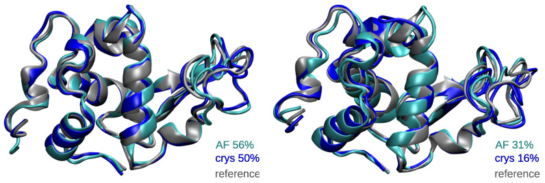
S-glutathionylation is an oxidative post-translational modification, which is involved in the regulation of many cell signaling pathways. Increasing amounts of studies show that it is crucial in cell homeostasis and deregulated in several pathologies. However, the effect of S-glutathionylation on proteins' structure and activity is poorly understood, and a drastic lack of structural information at the atomic scale remains. Studies based on the use of molecular dynamics simulations, which can provide important information about modification-induced modulation of proteins' structure and function, are also sparse, and there is no benchmarked force field parameters for this modified cysteine. In this contribution, we provide robust AMBER parameters for S-glutathionylation, which we tested extensively against experimental data through a total of 33 μs molecular dynamics simulations. We show that our parameter set efficiently describes the global and local structural properties of S-glutathionylated proteins. These data provide the community with an important tool to foster new investigations into the effect of S-glutathionylation on protein dynamics and function, in a common effort to unravel the structural mechanisms underlying its critical role in cellular processes.

Keywords: AMBER force field parameters; S-glutathionylation; molecular dynamics simulations; post-translational modifications; redox modifications.


Conflict of interest statement

The authors declare no conflict of interest.

Figures

Figure 1
Figure 1
(A) Structure of a glutathionylated cysteine. The glycine, cysteine, and glutamate residues within the glutathionylation moiety (SSG) are labeled in grey. The target cysteine backbone atoms are displayed as if embedded in a protein (without capping). (B) A few examples of proteins regulated by S-glutathionylation, with respect to their function [7,18,19].
Figure 2
Figure 2
Boxplots of the RMSD values in Å of each system, considering all atoms (top) or the residues’ backbone atoms (bottom). RMSD values for dimers are shown by monomer (Mono 1 or Mono 2). The black lines inside each boxplot display the median values. The average and standard deviation values are written above each box.
Figure 3
Figure 3
Representative structures of the two main conformations of hLyso as calculated from the cluster analysis of MD ensembles. The structures of the crystal reference and from the AF and crys simulations are displayed in gray, cyan, and blue, respectively. The percentage of occurrence of each cluster (primary on the left and secondary on the right) is also given.
Figure 4
Figure 4
Boxplots of the S-glutathionylated cysteine (CSG) RMSD values in Å for each system. RMSD values for dimers are shown by monomer (Mono 1 or Mono 2). The black lines inside each boxplot display the median values. Average and standard deviation values are written above each box.
Figure 5
Figure 5
Times series of CSG RMSD values in Å of the three replicates for AF simulations of AtGrxC5, hGSTO2, PtGSTL1, PtGSTL3, PtGrx12, PtGSTF1, ScGrx2, and TvGSTO2C, which show better trends. Plots for the dimeric systems (hGSTO2, PtGSTF1, and TvGSTO2C) are paired in the columns, with CSG1 and CSG2 referring to Monomers 1 and 2, respectively.
Figure 6
Figure 6
(A) Representative structures of the major conformations of the CSG in AtGrxC5, TvGSTO2C Monomer 1, PtGSTL3, hLyso, ScTrx1, and PtTrxL2.1 as calculated from the cluster analysis of MD ensembles. Structures of the crystal reference and from the AF and crys simulations are displayed in gray, cyan, and blue, respectively. The percentage of occurrence of the clusters is also given. (B) Times series of the CSG RMSD values in Å of the three replicates for the AF (left) and crys (right) simulations of hLyso, ScTrx1, and PtTrxL2.1.
Figure 7
Figure 7
(A) CSG structure, highlighting the side chain atoms used to monitor hydrogen bonds. (BD) Distribution of distances corresponding to the native interactions identified in the reference structures of hLyso, PtTrxL2.1, and ScTrx1, respectively. Both distributions for AF (cyan) and crys (blue) simulations are shown. Cation–π interactions involving Y63 in hLyso are monitored as distances between CSG.N1 and the center of mass of the aromatic ring heavy atoms of Y63 (Y63.Ph).

References

    1. Dalle-Donne I., Rossi R., Colombo G., Giustarini D., Milzani A. Protein S-glutathionylation: A regulatory device from bacteria to humans. Trends Biochem. Sci. 2009;34:85–96. doi: 10.1016/j.tibs.2008.11.002. - DOI - PubMed
    1. Filomeni G., Rotilio G., Ciriolo M.R. Cell signalling and the glutathione redox system. Biochem. Pharmacol. 2002;64:1057–1064. doi: 10.1016/S0006-2952(02)01176-0. - DOI - PubMed
    1. Anathy V., Roberson E.C., Guala A.S., Godburn K.E., Budd R.C., Janssen-Heininger Y.M. Redox-based regulation of apoptosis: S-glutathionylation as a regulatory mechanism to control cell death. Antioxid. Redox Signal. 2012;16:496–505. doi: 10.1089/ars.2011.4281. - DOI - PMC - PubMed
    1. García-Giménez J.L., Olaso G., Hake S.B., Bönisch C., Wiedemann S.M., Markovic J., Dasi F., Gimeno A., Pérez-Quilis C., Palacios O., et al. Histone h3 glutathionylation in proliferating mammalian cells destabilizes nucleosomal structure. Antioxid. Redox Signal. 2013;19:1305–1320. doi: 10.1089/ars.2012.5021. - DOI - PMC - PubMed
    1. Kalinina E.V., Novichkova M.D. S-Glutathionylation and S-Nitrosylation as Modulators of Redox-Dependent Processes in Cancer Cell. Biochemistry. 2023;88:924–943. doi: 10.1134/S0006297923070064. - DOI - PubMed

LinkOut - more resources