. 2023 Oct 9;24(19):15029.
doi: 10.3390/ijms241915029.

The MADF-BESS Protein CP60 Is Recruited to Insulators via CP190 and Has Redundant Functions in Drosophila

Affiliations

The MADF-BESS Protein CP60 Is Recruited to Insulators via CP190 and Has Redundant Functions in Drosophila

Larisa Melnikova et al. Int J Mol Sci. .

Abstract

Drosophila CP190 and CP60 are transcription factors that are associated with centrosomes during mitosis. CP190 is an essential transcription factor and preferentially binds to housekeeping gene promoters and insulators through interactions with architectural proteins, including Su(Hw) and dCTCF. CP60 belongs to a family of transcription factors that contain the N-terminal MADF domain and the C-terminal BESS domain, which is characterized by the ability to homodimerize. In this study, we show that the conserved CP60 region adjacent to MADF is responsible for interacting with CP190. In contrast to the well-characterized MADF-BESS transcriptional activator Adf-1, CP60 is recruited to most chromatin sites through its interaction with CP190, and the MADF domain is likely involved in protein-protein interactions but not in DNA binding. The deletion of the Map60 gene showed that CP60 is not an essential protein, despite the strong and ubiquitous expression of CP60 at all stages of Drosophila development. Although CP60 is a stable component of the Su(Hw) insulator complex, the inactivation of CP60 does not affect the enhancer-blocking activity of the Su(Hw)-dependent gypsy insulator. Overall, our results indicate that CP60 has an important but redundant function in transcriptional regulation as a partner of the CP190 protein.

Keywords: MADF-BESS transcriptional factors; Su(Hw); architectural C2H2 proteins; gypsy; housekeeping genes; insulator.


Conflict of interest statement

The authors declare no conflict of interest.

Figures

Figure 1
Figure 1
CP60 is a component of the Su(Hw) insulator complex: (A) Schematic presentation of the Su(Hw) insulator complex. The previously described interactions [58] between the components of the complex are shown. The CP190 domains are shown as yellow ovals, with four zinc fingers shown as yellow boxes; the Mod(mdg4)-67.2 domains are shown as green ovals; Su(Hw) domains are shown as lilac boxes. Domain abbreviations: CID—CP190 interacting domain; Ac—C-terminal acidic domain; Zn—zinc-finger domain; LZ—leucine zipper; BTB—BTB/POZ domain; Q—glutamine-rich region; DD—dimerization domain; FLYWCH—FLYWCH-type zinc finger; SID—Su(Hw) interacting domain; D—asparagine-rich domain; M—the microtubule- and centrosome-associated domain; E—glutamine-rich C-terminal domain. Bold capital letters indicate the Su(Hw) binding site. (B) Nuclear extract from Drosophila S2 cells was immunoprecipitated with antibodies against CP60, and the immunoprecipitates (IP) were analyzed via Western blotting for the presence of Su(Hw), Mod(mdg4)-67.2, and CP190 proteins. Input is the input fraction (1% of the lysate used for immunoprecipitation); output is the supernatant after immunoprecipitation; IP is the immunoprecipitate; PI is immunoprecipitation with nonspecific IgG. (C) Nuclear extract from Drosophila S2 cells was immunoprecipitated with antibodies against Su(Hw), Mod(mdg4)-67.2, or CP190, and the immunoprecipitates (IP) were analyzed via Western blotting for the presence of CP60 protein. The uncropped images for (B,C) are shown in Supplementary Materials Figure S2. (D) Identification of direct interactions between CP60 and other components of the Su(Hw) insulator complex using the yeast two-hybrid assay. The results are summarized in the table with + and—signs referring to a strong interaction or no interaction, respectively. Interactions with pGBT9 or pGAD424 vectors were used as the negative control. Interactions between CP190 and Mod(mdg4)-67.2 with Su(Hw) were used as a positive control.
Figure 2
Figure 2
CP190 is responsible for CP60 recruitment to the Su(Hw) chromatin sites: (A) Western blot analysis of RNAi efficiency; wt—S2 cells without treatment; proteins indicated below the line (RNAi) were knocked out by RNAi; antibodies for staining are listed on the left of the panel. Anti-tubulin antibodies (αTub) were used as a loading control. The uncropped images are shown in Supplementary Materials Figure S3. (BE) ChIP-qPCR analysis of binding (B) Su(Hw), (C) CP190, (D) CP60, and (E) Mod(mdg4)-67.2 proteins to the selected Su(Hw) sites in wild-type (wt) S2 cells and after RNAi inactivation of each protein. The ras64B coding region (Ras) was used as a control that does not contain Su(Hw) binding sites. IgG—immunoprecipitation with nonspecific IgG. The percentage recovery of immunoprecipitated DNA (Y axis) was calculated relative to the amount of input DNA. Error bars indicate SDs of quadruplicate PCR measurements from two independent biological samples of chromatin. Asterisks indicate significance levels: * p < 0.05 and ** p < 0.01 (Student’s t-test). Dots on the bar plots indicate the values of individual experiments.
Figure 3
Figure 3
Analysis of the in vitro association between Su(Hw) or CP60 and Su(Hw) motifs: (A) Schematic representation of the tested Su(Hw) binding sites. The red squares show the localization of the Su(Hw) motifs. (B) The Adf-1 binding site from bxd PRE and purified Adf-1 protein were used in EMSA as the positive control. DNA fragments without protein are marked with a minus (−) sign. The triangle represents a threefold increase in Adf-1 concentration from 0.05 µg to 0.15 µg. (C) EMSA of the binding of Su(Hw) (0.05 µg) and CP60 (0.05 µg) proteins to DNA fragments 50 A, 62 D, 66 E, and 87 E. The Adf-1 binding site was used as the negative control for the Su(Hw) or CP60 proteins. (D) EMSA of the binding of CP60 and Su(Hw) proteins to the gypsy insulator (gypsy) and two copies of the minimal 125 bp 1A2 region (1A2 125 × 2), and 250 bp (1A2 250) and 450 bp (1A2 450) regions of 1A2 insulator site. The amount of Su(Hw) protein was increased threefold from 0.05 µg to 0.15 µg. Other designations are as in (B). (E) Cooperation of Su(Hw), CP190, and CP60 in the binding to the gypsy insulator. The combination of proteins (0.05 µg) is indicated above in the panel. The absence of the corresponding protein in the EMSA probe is marked as a minus (−) sign. (F) EMSA supershift by CP60 antibodies and different combinations of Su(Hw) insulator proteins (0.05 µg). Ab—CP60 antibodies were added to the probe. Other designations as in (E).
Figure 4
Figure 4
Mapping domains of CP60 that are responsible for homodimerization and interaction with CP190 in the yeast two-hybrid assay. The scale at the top of the figure is in amino acids. A schematic representation of the CP60 protein is shown with deletion margins on the left. The MADF domain is shown as a green rectangle. Conserved regions 1–3 are represented by red rectangles. The fourth C-terminal conserved region is shown as a blue rectangle. The interactions are summarized in columns on the right-hand side with + and − referring to the presence and absence of interactions, respectively.
Figure 5
Figure 5
Multiple alignment analysis of the CP60 C-terminal conserved region (440–441 aa) sequence with different known BESS domains. Alignment regions are shown as colored boxes or outlines that enclose one or more residue symbols. The residue coloring corresponds to the Clustal X color code (Supplementary Table S1).
Figure 6
Figure 6
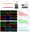
Functional analysis of the Map60 gene: (A) CRISPR/Cas9 deletion of the Map60 gene. The CP60 coding regions are shown as orange boxes. White rectangles represent the 5′ and 3′UTR. CRISPR targets are shown as vertical bars. Primer sequences used in the CRISPR/Cas9 genome editing are given in Supplementary Table S2. The Red reporter (magenta box), controlled by the 3xP3 promoter (black line), was used for the selection of the Map60 deletion. The attP and lox sites were used for genome manipulation and are shown as a green box and vertical yellow arrows, respectively. SV40 terminator is shown as a blue box. The two black arrows represent primers 1 and 2, which were used to test the obtained mutations using PCR. (B) PCR analysis of genomic DNA from Map60Δ1 lines after CRE/loxP excision of marker dsRed cassette (ΔdsRed). The molecular weight in bp is shown on the right. (C) Western blot analysis (8% SDS PAGE) of protein extracts prepared as described previously [72] from adult three-day-old males of the wild-type (wt) line and three lines homozygous for Map60Δ1. The membrane was sequentially stained with tested polyclonal rat antibodies against CP60 (αCP60) and antibodies against tubulin (αTub) as loading control. The molecular weight in kDa is shown on the left. The uncropped images are shown in Supplementary Materials Figure S7. (D) Polytene chromosomes from the salivary glands of third-instar y2w1118ct6 (wt) and y2w1118ct6; Map60Δ1-1/Map60Δ1-1 (Map60Δ1-1) larvae costained with rat anti-CP60 antibodies (green), rabbit anti-CP190 antibodies (red), and DAPI (blue) Scale bars, 10 μm. (E) ChIP-qPCR analysis of Su(Hw), CP190, and CP60 binding to Su(Hw) sites (description in Figure 2) in wt and Map60Δ1-1 (Δ1) lines. The ras64B coding region (Ras) that did not contain Su(Hw) binding sites was used as a control. IgG—immunoprecipitation with nonspecific IgG. The percentage recovery of immunoprecipitated DNA (Y axis) was calculated relative to the amount of input DNA. Error bars indicate SDs of quadruplicate PCR measurements from two independent biological samples of chromatin. Significance levels: p < 0.05 (Student’s t-test). Dots on the bar plots indicate the values of individual experiments.
Figure 7
Figure 7
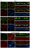
Testing the genome-wide distribution of CP60 and its colocalization with the components of the Su(Hw) complex. Polytene chromosomes from the salivary glands of third-instar (A) y2w1118ct6 (wt) and (B) y2w1118ct6; Cp1902/Cp1903 (Cp1902/Cp1903) larvae costained with rat anti-CP60 antibodies (green), rabbit anti-CP190 or anti-Su(Hw) antibodies (red), and DAPI (blue). Scale bars, 10 μm.
Figure 8
Figure 8
CP190 is responsible for CP60 recruitment to dCTCF/CP190 and BEAF/CP190 sites. ChIP-qPCR analysis of binding of dCTCF, CP190, and CP60 to the (A) dCTCF (Fab3, Fab4, Fab8, and MCP)- and Pita (Fab7)-dependent sites of the Bitorax complex. (B) BEAF-dependent sites from promoter regions of the aurora, cg3281, and janA genes. (C) Promoter and regulatory regions from the Bitorax complex are not bound by CP190 but are enriched in isoforms of the Mod(mdg4) protein. The ras64B coding region (Ras) was used as a control devoid of dCTCF/CP190- and BEAF/CP190 binding sites. IgG—immunoprecipitation with nonspecific IgG. The percentage recovery of immunoprecipitated DNA (Y axis) was calculated relative to the amount of input DNA. Error bars indicate SDs of quadruplicate PCR measurements from two independent biological samples of chromatin. Significance levels: p < 0.01 (Student’s t-test). Dots on the bar plots indicate the values of individual experiments.

Similar articles

Cited by

References

    1. Cavalheiro G.R., Pollex T., Furlong E.E. To Loop or Not to Loop: What Is the Role of TADs in Enhancer Function and Gene Regulation? Curr. Opin. Genet. Dev. 2021;67:119–129. doi: 10.1016/j.gde.2020.12.015. - DOI - PubMed
    1. Furlong E.E.M., Levine M. Developmental Enhancers and Chromosome Topology. Science. 2018;361:1341–1345. doi: 10.1126/science.aau0320. - DOI - PMC - PubMed
    1. Kim J., Dean A. Enhancers Navigate the Three-Dimensional Genome to Direct Cell Fate Decisions. Curr. Opin. Struct. Biol. 2021;71:101–109. doi: 10.1016/j.sbi.2021.06.005. - DOI - PubMed
    1. Razin S.V., Ulianov S.V., Iarovaia O.V. Enhancer Function in the 3D Genome. Genes. 2023;14:1277. doi: 10.3390/genes14061277. - DOI - PMC - PubMed
    1. da Costa-Nunes J.A., Noordermeer D. TADs: Dynamic Structures to Create Stable Regulatory Functions. Curr. Opin. Struct. Biol. 2023;81:102622. doi: 10.1016/j.sbi.2023.102622. - DOI - PubMed

MeSH terms

LinkOut - more resources