Review
. 2023 Sep 24;16(19):6381.
doi: 10.3390/ma16196381.

Metal Chalcogenide-Hydroxide Hybrids as an Emerging Family of Two-Dimensional Heterolayered Materials: An Early Review

Affiliations
Review

Metal Chalcogenide-Hydroxide Hybrids as an Emerging Family of Two-Dimensional Heterolayered Materials: An Early Review

Yuri Mikhlin et al. Materials (Basel). .

Abstract

Two-dimensional (2D) materials and phenomena attract huge attention in modern science. Herein, we introduce a family of layered materials inspired by the minerals valleriite and tochilinite, which are composed of alternating "incompatible", and often incommensurate, quasi-atomic sheets of transition metal chalcogenide (sulfides and selenides of Fe, Fe-Cu and other metals) and hydroxide of Mg, Al, Fe, Li, etc., stacked via electrostatic interaction rather than van der Waals forces. We survey the data available on the composition and structure of the layered minerals, laboratory syntheses of such materials and the effect of reaction conditions on the phase purity, morphology and composition of the products. The spectroscopic results (Mössbauer, X-ray photoelectron, X-ray absorption, Raman, UV-vis, etc.), physical (electron, magnetic, optical and some others) characteristics, a specificity of thermal behavior of the materials are discussed. The family of superconductors (FeSe)·(Li,Fe)(OH) having a similar layered structure is briefly considered too. Finally, promising research directions and applications of the valleriite-type substances as a new class of prospective multifunctional 2D materials are outlined.

Keywords: Mössbauer spectroscopy; XPS; heterostructure; hydrothermal synthesis; layered minerals; tochilinite; two-dimensional materials; valleriite.


Conflict of interest statement

The authors declare no conflict of interest. The funders had no role in the design of the study; in the collection, analyses, or interpretation of data; in the writing of the manuscript; or in the decision to publish the results.

Figures

Figure 1
Figure 1
(a) Number of publications in the domain of 2D nanomaterials [7] (licensed under CC BY 4.0). (b) Scheme of formation of heterostructured 2D materials [35]; permission from Springer Nature.
Figure 3
Figure 3
Schematic representation of (a) brucite Mg(OH)2 structure viewed down the z axis; (b) the tetragonal structure and XRD pattern of mackinawite FeS (synthetic). Reproduced from [54] with permission from APS.
Figure 6
Figure 6
(a) Structure of tochilinite, (b) coordination units FeS4 in tochilinite and VS6 in yushkinite, and (c) structure of vyalsovite [85], permission from Springer Publishing Company.
Figure 2
Figure 2
Typical crystal structures of layered clay minerals: (a) montmorillonite, a 2:1 type smectite, with layers consisting of two tetrahedral sheets and one octahedral sheet separated by the interlayer space with hydrated cations; (b) kaolinite, a 1:1 type clay mineral, with layers consisting of one tetrahedral and one octahedral sheet. Reproduced from Ref. [28] with permission from the Royal Society of Chemistry (RSC). (c) Schematic LDH structure [29]. © Distributed by Creative Commons Attribution 3.0 License.
Figure 4
Figure 4
Schemes of crystalline structures of (a,b) “single-layer” and (c) “three-layer” valleriites, and (d) coordination units in the hydroxide and sulfide layers.
Figure 5
Figure 5
SEM images of (a) tochilinite fibers from Cornwall, Pennsylvania [78] (permission from the Mineralogical Society of America), and (b) ferrotochilinite from Noril’sk, Russia [80] (the scale length is probably 40 μm but not 40 mm) (permission from Springer Nature).
Figure 7
Figure 7
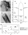
(a) Phase diagram showing hydrothermal formation of valleriite and other compounds, and (b,c) TEM micrographs of valleriite particles: (c) shows enlarged area A. Lines bounding the different phase regions were not determined accurately and are given as guides only [89]. Permission from Elsevier.
Figure 8
Figure 8
(a) Atomic structure of the Cu-Fe sulfide and (Mg,Fe,Al)(OH)2 sheets in valleriite (slightly tilted for better view), (b) X-ray diffraction patterns of valleriite samples synthesized a—without Al, b—with Al and c, d—various initial ratios of Fe, Cu, Mg precursors. XRD data in panel (c) illustrate the effect of addition of e, f—Cr, g—Co and h—La. Reflections of Mg and La hydroxides are marked as Mg (a) and La (h). TEM images (d,e,g,h) and particle size distributions (f,i) are given for the samples a and b, respectively [93]. Permission from RSC.
Figure 9
Figure 9
(a,b) TEM images of plate-like and tubular tochilinite particles prepared under hydrogen pressure and phase diagrams for the formation of (c) Mg,Fe-tochilinite [94] (permission from Elsevier) and (d) ferrotochilinite [95] during the interaction of H2S with metal hydroxides.
Figure 10
Figure 10
Upper panels: (a) SEM and (b) TEM images of Fe0.76S⋅0.86 [Fe2+0.01Fe3+0.56Mg2+0.43(OH)2.01] prepared using elemental Fe and S [101]. Lower panels: (c,d) typical TEM images, (e) electron diffractogram and (f) X-ray diffraction patterns of tochilinites prepared using atomic proportions of reagents Fe 2, Mg 1.5, S 15 (black), Fe 2, Mg 1.5, S 15, Al 0.5 (red), and Fe 2, Mg 1.5, S 15, Li 0.5 (green); asterisks designate reflections of brucite [103]. Reproduced with permission from RSC.
Figure 11
Figure 11
Left panels: (a) FESEM of [Li0.85Fe0.15OH][FeS] single crystals. (b) EDX results of the single crystals. (c) Elemental distribution of the Fe, O and S elements in the [Li0.85Fe0.15OH][FeS] single crystal. (d) X-ray diffraction of [Li0.85Fe0.15OH][FeS] [111] (permission from RSC). Right panels: (e) a schematic illustration of structural changes in the process of the hydrothermal ionic exchange reaction with the starting materials of big matrix crystals of K0.8Fe1.6Se2, LiOH·H2O, Fe and CH4N2Se. (f,g) The XRD patterns of (00l) type for the K0.8Fe1.6Se2 and the (Li0.84Fe0.16)OH·Fe0.98Se crystals, respectively, demonstrating their crystal orientations along (001) planes. The insets show the corresponding photographs of the crystals. Reproduced from [115] with permissions of APS.
Figure 12
Figure 12
Upper panels: X-ray photoelectron spectra, lower panels: Mössbauer spectra, temperature (FC and ZFC) and field dependences (hysteresis loops at 4.2 K) of magnetization and reciprocal susceptibility 1/χ of valleriite. The samples were synthesized with different proportions of Fe and Cu precursors without Al (a) and with Al (b), (c), and with Cr (d) [93]. Permission from RSC.
Figure 13
Figure 13
Left panels: The hydrodynamic diameters Dh (a,d), zeta potentials (b,e), and UV-vis-NIR absorption spectra (c,f) of valleriite hydrosols spontaneously formed during washing (upper panels (ac) and a zeta potential—pH plot for the sample b5 in an insert) and prepared by means of sonification of corresponding residues in aqueous 2 mM SDS solution (df). In upper panels, the samples were synthesized (32 h) using the initial precursor ratios: a3, a5—Al 0.5, Fe 2, Cu 2, Mg 2, S 14; b3, b5—Al 0, Fe 2, Cu 2, Mg 2, S 14, with indexes 3 and 5 standing for the number of the washing stage in which the particular sol was formed. In lower panels, hydrosol sample a contained no Al; b, c—Al-containing, e—Cr-doped [93]. Right panels. (g): UV-vis-NIR absorption spectra of aqueous colloids of tochilinite synthesized hydrothermally using the proportions of reagents a—Fe 2, Mg 1.5, S 15, b—Fe 2, Mg 1.5, S 15, Al 0.5, c—Fe 2, Mg 1.5, S 15, Li 0.5, d—Fe 2, Mg 1.5, S 15, Al 0.5, Li 0.5; and dispersed in water. (h): Simplified diagram of the electron levels for Fe2+ cations assuming tetrahedral coordination (the levels for Fe3+ are not shown), S ligands, and tentative optical charge-transfer transitions in the sulfide layer of tochilinite [103]. Permissions from RSC.
Figure 14
Figure 14
(a): Imaginary part of dielectric permittivity for tochilinites prepared with the atomic ratios of precursors a—Fe 2, Mg 2, S 15, b—Fe 2, Mg 2, S 15, Al 0.5, c—Fe 2, Mg 2, S 15, Li 0.5. (b): dielectric loss tangents at various frequencies as a function of temperature for the sample b; arrow marks increasing frequency. (c): peak frequency vs. reciprocal temperature (Arrhenius plot) for the sample b. Reproduced from Ref. [103] with permission from RSC.
Figure 15
Figure 15
TGA and DSC profiles acquired in Ar (left panels (a,b)) and 20% O2 + 80% N2 media (right panels (c,d)) for synthetic valleriite samples prepared with no additives (a,c) or using Al (b,d) as a modifier decreasing the content of Fe in hydroxide layers. Adapted from [136]. © Distributed by Creative Commons Attribution 3.0 License.

Similar articles

References

    1. Novoselov K.S., Geim A.K., Morozov S.V., Jiang D., Zhang Y., Dubonos S.V., Grigorieva I.V., Firsov A.A. Electric Field Effect in Atomically Thin Carbon Films. Science. 2004;306:67–69. doi: 10.1126/science.1102896. - DOI - PubMed
    1. Geim A.K. Graphene: Status and Prospects. Science. 2009;324:1530–1534. doi: 10.1126/science.1158877. - DOI - PubMed
    1. Urade A.R., Lahiri I., Suresh K.S. Graphene Properties, Synthesis and Applications: A Review. JOM. 2023;75:614–630. doi: 10.1007/s11837-022-05505-8. - DOI - PMC - PubMed
    1. Razaq A., Bibi F., Zheng X., Papadakis R., Jafri S.H.M., Li H. Review on Graphene-, Graphene Oxide-, Reduced Graphene Oxide-Based Flexible Composites: From Fabrication to Applications. Materials. 2022;15:1012. doi: 10.3390/ma15031012. - DOI - PMC - PubMed
    1. Xia Y., Gao W., Gao C. A Review on Graphene-Based Electromagnetic Functional Materials: Electromagnetic Wave Shielding and Absorption. Adv. Funct. Mater. 2022;32:2204591. doi: 10.1002/adfm.202204591. - DOI

LinkOut - more resources