

Influence of Aortic Valve Stenosis and Wall Shear Stress on Platelets Function
- PMID: 37834945
- PMCID: PMC10573628
- DOI: 10.3390/jcm12196301
Influence of Aortic Valve Stenosis and Wall Shear Stress on Platelets Function
Abstract
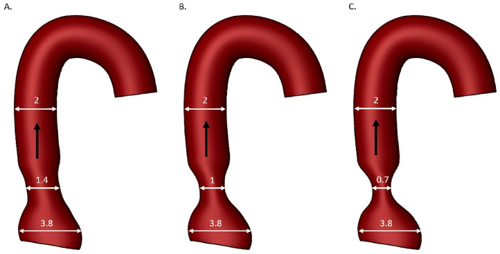
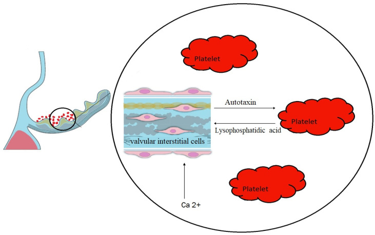
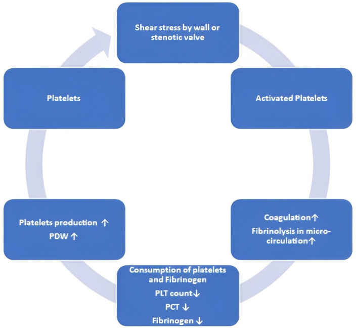
Aortic valve stenosis (AS) is a common heart valve disease in the elderly population, and its pathogenesis remains an interesting area of research. The degeneration of the aortic valve leaflets gradually progresses to valve sclerosis. The advanced phase is marked by the presence of extracellular fibrosis and calcification. Turbulent, accelerated blood flow generated by the stenotic valve causes excessive damage to the aortic wall. Elevated shear stress due to AS leads to the degradation of high-molecular weight multimers of von Willebrand factor, which may involve bleeding in the mucosal tissues. Conversely, elevated shear stress has been associated with the release of thrombin and the activation of platelets, even in individuals with acquired von Willebrand syndrome. Moreover, turbulent blood flow in the aorta may activate the endothelium and promote platelet adhesion and activation on the aortic valve surface. Platelets release a wide range of mediators, including lysophosphatidic acid, which have pro-osteogenic effects in AS. All of these interactions result in blood coagulation, fibrinolysis, and the hemostatic process. This review summarizes the current knowledge on high shear stress-induced hemostatic disorders, the influence of AS on platelets and antiplatelet therapy.
Keywords: antiplatelet therapy; aortic stenosis; cerebrovascular events; hemodynamics; platelets function; valvular heart disease; wall shear stress.
Conflict of interest statement
The authors declare no conflict of interest.
Figures
References
-
- Iung B., Baron G., Butchart E.G., Delahaye F., Gohlke-Bärwolf C., Levang O.W., Tornos P., Vanoverschelde J.-L., Vermeer F., Boersma E., et al. A prospective survey of patients with valvular heart disease in Europe: The Euro Heart Survey on Valvular Heart Disease. Eur. Heart J. 2003;24:1231–1243. doi: 10.1016/S0195-668X(03)00201-X. - DOI - PubMed
-
- Bouchareb R., Boulanger M.-C., Tastet L., Mkannez G., Nsaibia M.J., Hadji F., Dahou A., Messadeq Y., Arsenault B.J., Pibarot P., et al. Activated platelets promote an osteogenic programme and the progression of calcific aortic valve stenosis. Eur. Heart J. 2019;40:1362–1373. doi: 10.1093/eurheartj/ehy696. - DOI - PMC - PubMed
Publication types
LinkOut - more resources
Full Text Sources
Research Materials
