. 2023 Sep 27;15(19):4748.
doi: 10.3390/cancers15194748.

Evaluation of Prognostic Parameters to Identify Aggressive Penile Carcinomas

Affiliations

Evaluation of Prognostic Parameters to Identify Aggressive Penile Carcinomas

Jan Niklas Mink et al. Cancers (Basel). .

Abstract

Background: Advanced penile carcinoma is characterized by poor prognosis. Most data on prognostic factors are based on small study cohorts, and even meta-analyses are limited in patient numbers. Therefore, there is still a lack of evidence for clinical decisions. In addition, the most recent TNM classification is questionable; in line with previous studies, we found that it has not improved prognosis estimation.

Methods: We evaluated 297 patients from Germany, Russia, and Portugal. Tissue samples from 233 patients were re-analyzed by two experienced pathologists. HPV status, p16, and histopathological parameters were evaluated for all patients.

Results: Advanced lymph node metastases (N2, N3) were highly significantly associated with reductions in metastasis-free (MFS), cancer-specific (CS), and overall survival (OS) rates (p = <0.001), while lymphovascular invasion was a significant parameter for reduced CS and OS (p = 0.005; p = 0.007). Concerning the primary tumor stage, a significant difference in MFS was found only between pT1b and pT1a (p = 0.017), whereas CS and OS did not significantly differ between T categories. In patients without lymph node metastasis at the time of primary diagnosis, lymphovascular invasion was a significant prognostic parameter for lower MFS (p = 0.032). Histological subtypes differed in prognosis, with the worst outcome in basaloid carcinomas, but without statistical significance. HPV status was not associated with prognosis, either in the total cohort or in the usual type alone.

Conclusion: Lymphatic involvement has the highest impact on prognosis in penile cancer, whereas HPV status alone is not suitable as a prognostic parameter. The pT1b stage, which includes grading, as well as lymphovascular and perineural invasion in the T stage, seems questionable; a revision of the TNM classification is therefore required.

Keywords: HPV; histological subtype; p16; penile cancer; prognosis.


Conflict of interest statement

The authors declare no conflict of interest.

Figures

Figure 1
Figure 1
(a) Kaplan–Meier curve of MFS related to histological subtypes. (b) Kaplan–Meier curve of CSS related to histological subtypes. (c) Kaplan–Meier curve of OS related to histological subtypes.
Figure 1
Figure 1
(a) Kaplan–Meier curve of MFS related to histological subtypes. (b) Kaplan–Meier curve of CSS related to histological subtypes. (c) Kaplan–Meier curve of OS related to histological subtypes.
Figure 2
Figure 2
(a) Kaplan–Meier curve of MFS related to HPV status. (b) Kaplan–Meier curve of CSS related to HPV status. (c) Kaplan–Meier curve of OS related to HPV status.
Figure 2
Figure 2
(a) Kaplan–Meier curve of MFS related to HPV status. (b) Kaplan–Meier curve of CSS related to HPV status. (c) Kaplan–Meier curve of OS related to HPV status.
Figure 3
Figure 3
(a) Kaplan–Meier curve of MFS related to HPV status in the usual type. (b) Kaplan–Meier curve of CSS related to HPV status in the usual type. (c) Kaplan–Meier curve of OS related to HPV status in the usual type.
Figure 3
Figure 3
(a) Kaplan–Meier curve of MFS related to HPV status in the usual type. (b) Kaplan–Meier curve of CSS related to HPV status in the usual type. (c) Kaplan–Meier curve of OS related to HPV status in the usual type.
Figure 4
Figure 4
(a) Kaplan–Meier curve of MFS related to the T stage. (b) Kaplan–Meier curve of CSS related to T stage. (c) Kaplan–Meier curve of OS related to T stage.
Figure 4
Figure 4
(a) Kaplan–Meier curve of MFS related to the T stage. (b) Kaplan–Meier curve of CSS related to T stage. (c) Kaplan–Meier curve of OS related to T stage.
Figure 5
Figure 5
(a) Kaplan–Meier curve of MFS related to nodal status. (b) Kaplan–Meier curve of CSS related to nodal status. (c) Kaplan–Meier curve of OS related to nodal status.
Figure 5
Figure 5
(a) Kaplan–Meier curve of MFS related to nodal status. (b) Kaplan–Meier curve of CSS related to nodal status. (c) Kaplan–Meier curve of OS related to nodal status.
Figure 6
Figure 6
(a) Kaplan–Meier curve of MFS related to grading. (b) Kaplan–Meier curve of CSS related to grading. (c) Kaplan–Meier curve of OS related to grading.
Figure 6
Figure 6
(a) Kaplan–Meier curve of MFS related to grading. (b) Kaplan–Meier curve of CSS related to grading. (c) Kaplan–Meier curve of OS related to grading.
Figure 7
Figure 7
(a) Kaplan–Meier curve of MFS related to vascular invasion. (b) Kaplan–Meier curve of CSS related to vascular invasion. (c) Kaplan–Meier curve of OS related to vascular invasion.
Figure 7
Figure 7
(a) Kaplan–Meier curve of MFS related to vascular invasion. (b) Kaplan–Meier curve of CSS related to vascular invasion. (c) Kaplan–Meier curve of OS related to vascular invasion.
Figure 8
Figure 8
(a) Kaplan–Meier curve of MFS related to lymphovascular invasion. (b) Kaplan–Meier curve of CSS related to lymphovascular invasion. (c) Kaplan–Meier curve of OS related to lymphovascular invasion.
Figure 8
Figure 8
(a) Kaplan–Meier curve of MFS related to lymphovascular invasion. (b) Kaplan–Meier curve of CSS related to lymphovascular invasion. (c) Kaplan–Meier curve of OS related to lymphovascular invasion.
Figure 9
Figure 9
(a) Kaplan–Meier curve of MFS related to perineural invasion. (b) Kaplan–Meier curve of CSS related to perineural invasion. (c) Kaplan–Meier curve of OS related to perineural invasion.
Figure 9
Figure 9
(a) Kaplan–Meier curve of MFS related to perineural invasion. (b) Kaplan–Meier curve of CSS related to perineural invasion. (c) Kaplan–Meier curve of OS related to perineural invasion.
Figure 10
Figure 10
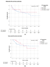
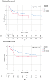
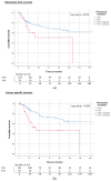
(a) Kaplan–Meier curve of MFS related to lymphovascular invasion in node-negative patients. (b) Kaplan–Meier curve of CSS related to lymphovascular invasion in node-negative patients. (c) Kaplan–Meier curve of OS related to lymphovascular invasion in node-negative patients.
Figure 10
Figure 10
(a) Kaplan–Meier curve of MFS related to lymphovascular invasion in node-negative patients. (b) Kaplan–Meier curve of CSS related to lymphovascular invasion in node-negative patients. (c) Kaplan–Meier curve of OS related to lymphovascular invasion in node-negative patients.
Figure 10
Figure 10
(a) Kaplan–Meier curve of MFS related to lymphovascular invasion in node-negative patients. (b) Kaplan–Meier curve of CSS related to lymphovascular invasion in node-negative patients. (c) Kaplan–Meier curve of OS related to lymphovascular invasion in node-negative patients.

References

    1. Bleeker M.C.G., Heideman D.A.M., Snijders P.J.F., Horenblas S., Dillner J., Meijer C.J.L.M. Penile Cancer: Epidemiology, Pathogenesis and Prevention. World J. Urol. 2009;27:141–150. doi: 10.1007/s00345-008-0302-z. - DOI - PubMed
    1. Fu L., Tian T., Yao K., Chen X.-F., Luo G., Gao Y., Lin Y.-F., Wang B., Sun Y., Zheng W., et al. Global Pattern and Trends in Penile Cancer Incidence: Population-Based Study. JMIR Public Health Surveill. 2022;8:e34874. doi: 10.2196/34874. - DOI - PMC - PubMed
    1. Thomas A., Necchi A., Muneer A., Tobias-Machado M., Tran A.T.H., Van Rompuy A.-S., Spiess P.E., Albersen M. Penile Cancer. Nat. Rev. Dis. Primers. 2021;7:11. doi: 10.1038/s41572-021-00246-5. - DOI - PubMed
    1. Christodoulidou M., Sahdev V., Houssein S., Muneer A. Epidemiology of Penile Cancer. Curr. Probl. Cancer. 2015;39:126–136. doi: 10.1016/j.currproblcancer.2015.03.010. - DOI - PubMed
    1. Kayes O., Ahmed H.U., Arya M., Minhas S. Molecular and Genetic Pathways in Penile Cancer. Lancet Oncol. 2007;8:420–429. doi: 10.1016/S1470-2045(07)70137-7. - DOI - PubMed

LinkOut - more resources