. 2023 Oct 4;15(19):4849.
doi: 10.3390/cancers15194849.

Mistletoe Extracts from Different Host Trees Disparately Inhibit Bladder Cancer Cell Growth and Proliferation

Affiliations

Mistletoe Extracts from Different Host Trees Disparately Inhibit Bladder Cancer Cell Growth and Proliferation

Eva Juengel et al. Cancers (Basel). .

Abstract

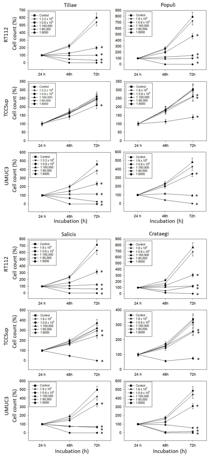
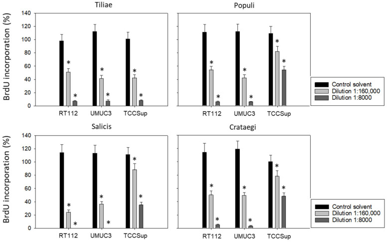
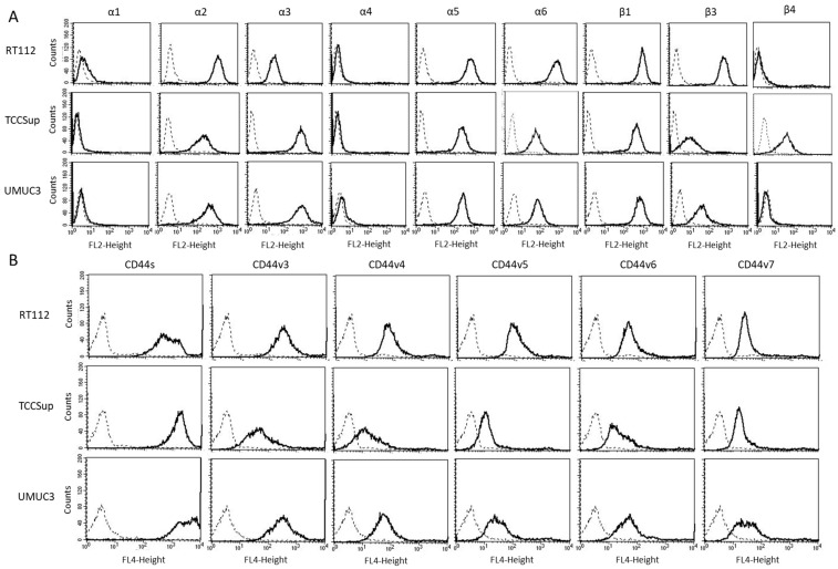
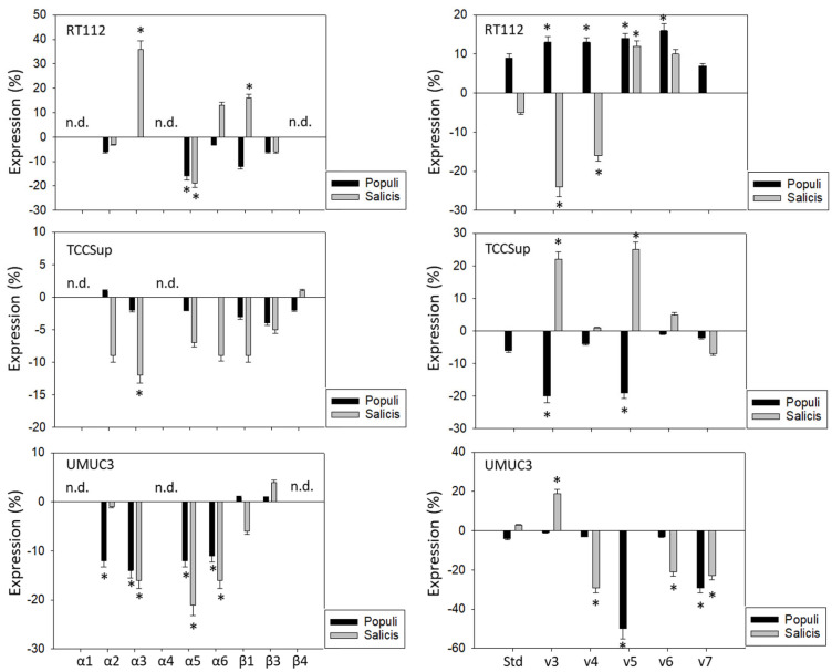
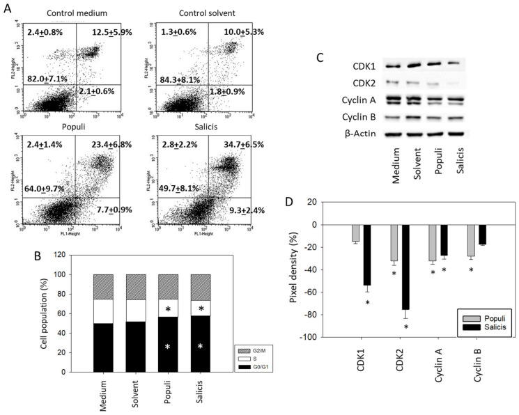
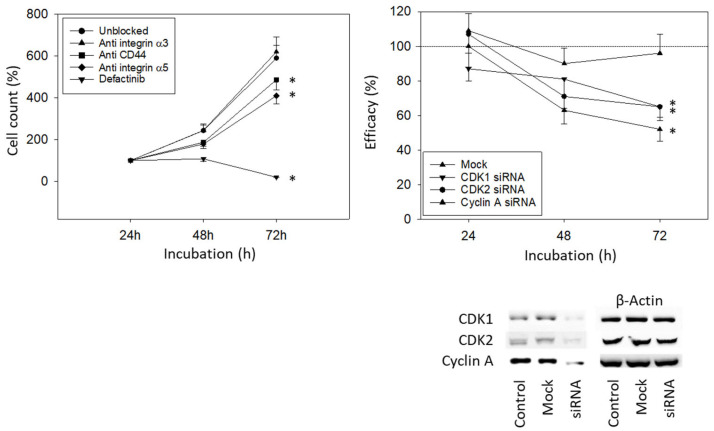
Extracts of European mistletoe (Viscum album) are popular as a complementary treatment for patients with many different cancer types. However, whether these extracts actually block bladder cancer progression remains unknown. The influence of different mistletoe extracts on bladder cancer cell growth and proliferation was investigated by exposing RT112, UMUC3, and TCCSup cells to mistletoe from hawthorn (Crataegi), lime trees (Tiliae), willow trees (Salicis), or poplar trees (Populi). The tumor cell growth and proliferation, apoptosis induction, and cell cycle progression were then evaluated. Alterations in integrin α and β subtype expression as well as CD44 standard (CD44s) and CD44 variant (CD44v) expressions were evaluated. Cell cycle-regulating proteins (CDK1 and 2, Cyclin A and B) were also investigated. Blocking and knock-down studies served to correlate protein alterations with cell growth. All extracts significantly down-regulated the growth and proliferation of all bladder cancer cell lines, most strongly in RT112 and UMUC3 cells. Alterations in CD44 expression were not homogeneous but rather depended on the extract and the cell line. Integrin α3 was, likewise, differently modified. Integrin α5 was diminished in RT112 and UMUC3 cells (significantly) and TCCSup (trend) by Populi and Salicis. Populi and Salicis arrested UMUC3 in G0/G1 to a similar extent, whereas apoptosis was induced most efficiently by Salicis. Examination of cell cycle-regulating proteins revealed down-regulation of CDK1 and 2 and Cyclin A by Salicis but down-regulation of CDK2 and Cyclin A by Populi. Blocking and knock-down studies pointed to the influence of integrin α5, CD44, and the Cyclin-CDK axis in regulating bladder cancer growth. Mistletoe extracts do block bladder cancer growth in vitro, with the molecular action differing according to the cell line and the host tree of the mistletoe. Integrating mistletoe into a guideline-based treatment regimen might optimize bladder cancer therapy.

Keywords: bladder cancer; cell cycling; growth; mistletoe; proliferation.


Conflict of interest statement

The authors declare no conflict of interest and that the sponsors had no role in data collection, analysis and interpretation. The funders had no input in study design; in the collection, analyses, discussion or interpretation of data; in the writing of the manuscript, or in the decision to publish the results, and did not discuss the manuscript or the data.

Figures

Figure 1
Figure 1
Influence of increasing concentrations of four mistletoe extracts (Tiliae, Populi, Salicis, Crataegi) on the growth of RT112, TCCSup, and UMUC3 bladder cancer cell lines. The cell number was evaluated after 24, 48, and 72 h by the MTT assay, whereby 24 h values were set to 100%. “Control” indicates medium control. Error bars indicate standard deviation. Experiments were performed three times. * indicates significant difference to the corresponding control.
Figure 2
Figure 2
Influence of four mistletoe extracts (Tiliae, Populi, Salicis, Crataegi) on the proliferation of RT112, UMUC3, and TCCSup cells. Proliferative activity was analyzed by the BrdU incorporation assay following 48 h mistletoe incubation. Error bars indicate standard deviation. * indicates a significant difference to controls (tumor cells exposed to culture medium alone) set to 100%. n = 3.
Figure 3
Figure 3
(A) surface expression of α and β integrins. (B) surface expression of CD44 standard (CD44s) and CD44 variants v3, v4, v5, v6, and v7. The abscissa shows the relative logarithmic distribution of the relative fluorescence intensity, the ordinate shows cell number. The solid line indicates specific fluorescence, the dashed shows the IgG1 isotype control. One representative figure from n = 3.
Figure 4
Figure 4
Influence of Populi and Salicis mistletoe extracts (Dilution 1:160,000) on the integrin (left) or CD44 standard (Std) and CD44 variants v3–v7 expression profile (right) of RT112, TCCSup, and UMUC3 cells. The untreated control is set to 0. Values are means ± SD, n = 4. Error bars indicate standard deviation. * indicates significant difference to controls. n.d.= not detectable.
Figure 5
Figure 5
Apoptosis and cell cycling. UMUC3 cells were treated with mistletoe from Populi or Salicis. Controls remained untreated (Medium) or were treated with solvent alone (Solvent). (A) apoptosis induction (dilution of 1:8000). Upper left quadrants show the percentage of cells in necrosis, upper right quadrants in late apoptosis, lower right quadrants in early apoptosis, and lower left quadrants vital cells (percentage values are means ± SD from n = 3). (B) cell cycle distribution evaluated after 24 h incubation. * indicates significant difference to the controls. (C) CDK1, CDK2, Cyclin A, and Cyclin B protein expression in UMUC3 cells detected by Western blotting (Full pictures of the Western blots are presented in the Supplementary Materials file). (D) presentation of the pixel density data. The results are given in percentage and related to CDK1, CDK2, Cyclin A, and Cyclin B expression in the untreated controls. (BD) depict a dilution of 1:160,000. * indicates significant difference to controls (n = 3).
Figure 6
Figure 6
Blocking and knock-down studies. (Left) UMUC3 cell number in response to integrin blockade (Anti integrin α3, anti integrin α5), to CD44s blockade (Anti CD44), and to blockade with defactinib (Defactinib). Untreated cells served as controls. Cell number was evaluated after 24, 48, and 72 h by the MTT assay (n = 3). (Right) cell growth of UMUC3 treated with a CDK1-, CDK2-, or Cyclin A-specific siRNA, evaluated by the MTT assay. The Western blot on lower right depicts reduction in CDK1 and CDK2 by siRNA knock down (Supplementary Figure S1). One representative of three separate experiments is shown. All values are related to the untreated controls set to 100%. Mock indicates scrambled controls. * indicates significant difference to the untreated (unblocked) control or to cells not treated with siRNA.

References

    1. Patel V.G., Oh W.K., Galsky M.D. Treatment of muscle-invasive and advanced bladder cancer in 2020. CA A Cancer J. Clin. 2020;70:404–423. doi: 10.3322/caac.21631. - DOI - PubMed
    1. Bilim V., Kuroki H., Shirono Y., Murata M., Hiruma K., Tomita Y. Advanced Bladder Cancer: Changing the Treatment Landscape. J. Pers. Med. 2022;12:1745. doi: 10.3390/jpm12101745. - DOI - PMC - PubMed
    1. Cockrell D.C., Rose T.L. Current Status of Perioperative Therapy in Muscle-Invasive Bladder Cancer and Future Directions. Curr. Oncol. Rep. 2023;25:511–520. doi: 10.1007/s11912-023-01390-9. - DOI - PubMed
    1. Lopez-Beltran A., Cimadamore A., Blanca A., Massari F., Vau N., Scarpelli M., Cheng L., Montironi R. Immune Checkpoint Inhibitors for the Treatment of Bladder Cancer. Cancers. 2021;13:131. doi: 10.3390/cancers13010131. - DOI - PMC - PubMed
    1. Song S., Cohen A.J., Lui H., Mmonu N.A., Brody H., Patino G., Liaw A., Butler C., Fergus K.B., Mena J., et al. Use of GoFundMe® to crowdfund complementary and alternative medicine treatments for cancer. J. Cancer Res. Clin. Oncol. 2020;146:1857–1865. doi: 10.1007/s00432-020-03191-0. - DOI - PMC - PubMed